More from the River Ehen

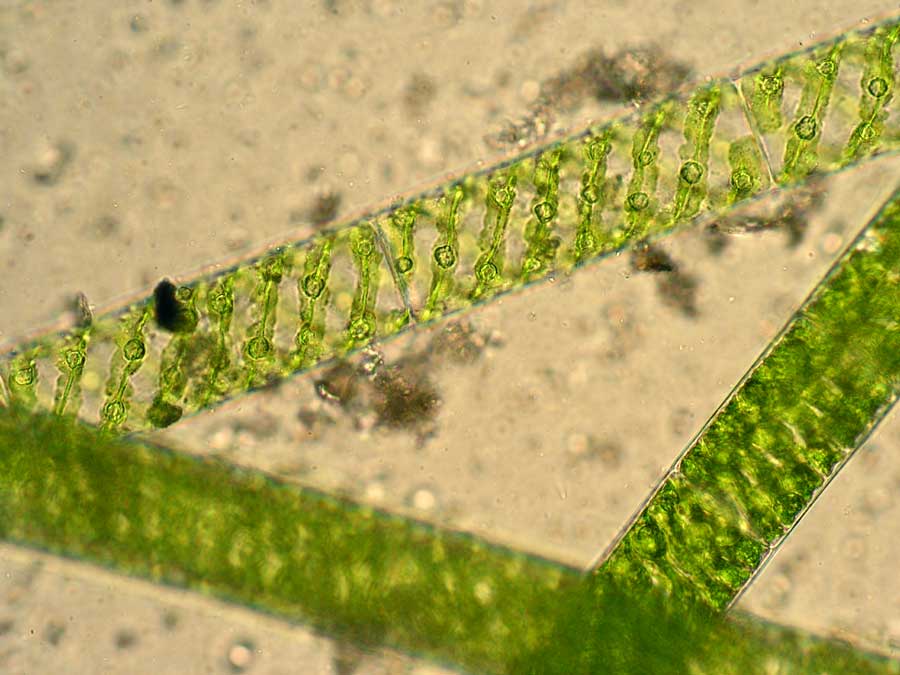
There were other colours of algae in the River Ehen too.  Further downstream, several of the stones had a reddish-pink hue which turned out to be composed of a completely different alga.   This was Audouinella hermanii, one of the relatively few freshwater species of red algae, more commonly encountered as seaweeds.   Like the green algae described in the previous post, this one is also composed of filaments although this time they have a red- rather than green colour.   There are some short side-branches bearing clusters of egg-shaped “carpogonia” (the female reproductive organs) and others which bear, like Draparnaldia, long, colourless hairs.  These, too, probably have a role in the phosphorus nutrition of the alga.

Ehen_130222_19Ehen_130222_7

The left hand image shows a cobble covered with Audouinella from the River Ehen, whilst the right hand image shows filaments and carpogonia under the microscope.  The filaments are approximately 10 micrometres (a hundredth of a millimetre) in diameter.

Some of the filaments also had short filaments of a blue-green alga, called Heteroleibleinia (formerly Lyngbya) rigidula growing on them and there were also many diatoms, particularly a species of Gomphonema growing on long stalks in and around the Audouinella filaments.   Later in the year, these growths of Audouinella get so completely smothered by diatoms that they lose their reddish appearance and become dark brown.

Ehen_130222_9Gomphonema_130222

The left hand image shows filaments of Heteroleibleinia rigidula growing on the Audouinella filaments; the right hand image shows four cells of the Gomphonema species (three in valve view and one, on the right, in girdle view).

The final image is a diorama putting all of these components together, with four distinct groups of algae in close juxtaposition.  The Audouinella filaments dominate the view but there are three diatoms, on their long stalks on the left hand side, along with a few filaments of Heteroleibleinia rigidula.  In the bottom right corner there are a few cells of Stigeoclonium tenue, a relative of Draparnaldia that I also saw growing amidst the Audouinella filaments.  Several of the Audouinella filaments end in colourless hairs on the right hand side.  The colour of the Audouinella reflects the pigment composition of the cells: the green of the chlorophyll is masked by two other pigments: phycocyanin, which is blue, and phycoerythrin, which is red.  A mix of Hooker’s Green, Ultramarine and Crimson Alizarin gives an approximation of the hue.  There should be several ribbon-shaped chloroplasts lying just inside the cell wall, but these were hard to resolve with a light microscope.

Ehen_130227

A three-dimensional representation of the Audouinella-dominated community in the River Ehen, February 2013.

Brian Whitton identified the Heteroleibleinia rigidula for me.

The River Ehen in February

We have had over a week of dry weather in the north of England, so the water level in the River Ehen was lower on Friday than I’ve seen it for a long time, although the weather was still cold enough for the occasional flurry of snow.   After my last experience plunging my arm into the river and soaking the sleeve of my shirt, I had come prepared today, with a box of vet’s insemination gloves which, though they don’t make the water any less cold, do at least keep my clothes dry.

Ehen_130222_3

Even though the trees overhanging the river were bare, the algae on the submerged stones were thriving, and added unexpected dashes of colour to the river bed.   Just downstream from the outflow from Ennerdale Water, the top surface of almost all the cobbles had a vivid green colour.  Closer examination showed a layer of fine green slimy filaments over a lower, browner and grittier layer.   Under the microscope, the green slimy filaments have the spiral chloroplasts characteristic of Spirogyra, albeit a different species to the one I found at Cassop (see post).   The lower layer was a mixture of tiny sediment particles and diatoms, mostly Tabellaria flocculosa and species of Gomphonema on long stalks.  Other diatoms included Achnanthidium minutissimum growing on short stalks themselves attached to the Gomphonema stalks, some fine needle-like cells of Fragilaria rumpens, also apparently attached to the stalks, and some cells of Brachysira amidst the humic particles trapped within the matrix.

Ehen_130222_1Ehen_130222_13

Spirogyra in the River Ehen: the image on the left shows the algae smothering a cobble; the image on the right is a microscopic view.  The filaments are 60 micrometres in diameter (= 0.06 mm).

The overall effect is of a microscopic “forest”, in which the Spirogyra forms a “canopy” and the diatoms the “understory” and “herbs”.   The mode of attachment of the Spirogyra was not evident, and Floras are vague on the subject, as Spirogyra is more often found as free-floating masses.  There is a passing reference to rhizoids in the Freshwater Algal Flora but no illustrations, so my illustration is based instead on a paper by Nagata (Plant Physiology 59: 680-683, 1977) which suggests that terminal cells secrete “sticky” substances which promote adhesion onto surfaces.  (Note, too, that the illustration is based on samples collected in October 2012, when the dominant Gomphonema species was G. acuminatum, whereas in February it was a species resembling G. clavatum).

Ehen_121011Ehen_121230

The Spirogyra-dominated community of the River Ehen: a two-dimensional illustration on the left, showing the Spirogyra “canopy” and the diatom-dominated understory (Gomphonema on stalks and zig-zag colonies of Tabellaria), and a three-dimensional diorama on the right.

This community had been present on most of my visits over the previous year. However, in February, there was also a green jelly-like growth on and around the same stones.  Under the microscope, this nondescript growth was transformed into beautiful growths of another green alga, Draparnaldia glomerata.   As for Spirogyra, this was composed of filaments of cells, though this time the chloroplast is not a spiral and, more significantly, the filaments are branched.   There is a main axis of cells, with clusters of side branches occurring at intervals along this.   These side branches each branch again, with the final branches terminating in long, colourless “hairs”.

Ehen_130222_15Ehen_130222_12

Draparnaldia glomerata from the River Ehen: left hand image shows a macroscopic view; right hand image shows the same organism but viewed at 400x magnification.  The main filament is 50 micrometres (one twentieth of a millimetre) in diameter.

Draparnaldia is adapted to living in situations where the nutrient concentrations in the water are generally low.  The hairs secrete an enzyme which enables Draparnaldia to obtain phosphorus from organic substances in the water.  The peat in the fells surrounding Ennerdale will provide plenty of this, particularly in the winter when rainfall and erosion will be at their peak.  Once these natural nutrients are supplemented by dissolved phosphorus in wastewater and from agricultural runoff, Draparnaldia loses its competitive edge and disappears.  The presence of so much Draparnaldia in the Ehen is, therefore, a clue to the generally good health of the river.

Whatever happened to the precautionary principle?

Something does not feel right when a group of ecologists gather on a concrete campus in the middle of a city, especially when we’re talking about the subject we’re talking about is restoration of rivers and lakes to their near-pristine condition (see Constable, Turner, Gainsborough post).   Aston University gives us a little inspiration by providing a muddy ornamental pond, just large enough to justify naming their conference suite “the Lakeside Centre”.

Aston_University

One other reflection from the meeting is how little reference official literature now makes to the Precautionary Principle.   This states that where there are threats of serious or irreversible environmental damage, lack of full scientific certainty should not be used as a reason for postponing cost effective measures to prevent environmental degradation.  It is enshrined in Article 191 of the Treaty on the Functioning of the European Union (follow link) but somewhere on the road between theory and practice high ideals collide with reality.  In some cases the Precautionary Principle even seems to be inverted.   The irony is that it is precisely those areas where scientific data do not permit a complete evaluation of risk that regulators are likely to prevaricate.  Environmental regulation involves “carrots” and “sticks”, with criminal prosecution as a last resort.   And the foundation of criminal law is the principle of “beyond reasonable doubt”, which does not sit comfortably with the Precautionary Principle.   Moreover, most regulation places if not a cost on a business then, at the very least, a limit on profitability.   The correlations ecologists find in their noisy datasets don’t necessarily translate into causation and, maybe, it is wrong to put these constraints on businesses without stronger certainty than most readings of the Precautionary Principle imply?

Another jargon phrase much loved by modern governments is “evidence-led policy” – again, a principle that is impossible to argue against yet, at the same time, contradictory, in some respects, to the Precautionary Principle.   Somewhere between having enough of a hunch to invoke the Precautionary Principle and enough certainty to satisfy a ministerial edict on evidence-led policy, most public servants (or, more likely, committees of public servants) will get cold feet.

Perhaps they are right?   I’ll finish back at the Lakeside Centre at Aston University where I am thinking through the implications of tighter standards for rivers, in particular.   Environmental regulation does not exist in isolation: they translate into the “sewerage charges” you’ll find on your next water bill.   The tight standards required for the Water Framework Directive means that these costs will creep upwards over the next few years.   The least we can do is make sure that you’ll see some benefits as a result.

Constable, Gainsborough, Turner …

If you went to the doctor with an ailment, your discussions would be based on a shared assumption about the properties of a healthy human body.  Understand this and you can put your ailment into perspective and, more importantly, the doctor knows how to treat you.  The same type of thinking is now permeating the world of ecology: if we all understand the characteristics of ecosystems in their natural state, then we have a basis for discussing what needs to be done (if anything) to restore those damaged by man’s actions.

One way of finding healthy ecosystems against which we can compare modern lakes and rivers is to look at evidence from the past, and there is a thriving discipline called palaeolimnology which addresses this question.   One of the uses of this is to find the dates before which the hand of man has little or no discernible effect on freshwater ecosystems.  Putting evidence from a very large number of studies together, colleagues at University College London suggested that the period between 1800 and 1850 could act as a rough “rule of thumb” for this baseline (see abstract).   There are a lot of caveats but , very roughly, the types of organisms we find preserved in lake sediments dated to before these dates correspond to the types of organisms we find in the most remote (and, therefore, pristine) lakes in Europe today.

I was thinking about this whilst visiting the Royal Academy’s exhibition on the origins of British landscape painting and, in particular, whilst standing in front of two large canvases by John Constable.  Both date from the 1830s, so lie within this period when we would expect rivers and lakes to be closer to their pristine states.   The problem is that Constable has depicted landscapes where the hand of man is very evident: there are mills and boatyards as well as artificially managed rivers and thriving agriculture.   You can see similar trends in prints based on Turner’s sketches: indeed, I can take you to a point on the River Tees near Barnard Castle where Turner clearly depicts a mill and a weir that have both now disappeared completely.   What is going on?

Constable and Turner both depict what are, to modern eyes, rural idylls.  They tie in with the 1800-1850 baseline insofar as they reflect a period in Britain before the use of fossil fuels became widespread, before sewage was routinely dumped in rivers and well before artificial fertilisers were widespread in agriculture.   So the chances are that the ecology in the rivers at the time was much closer to its natural state than we would find in many modern rivers.   But these were no pristine wildernesses: the Romantic painters were portraying landscapes where men were present but as likely to be dominated by nature as to dominate it themselves.  Their paintings suggest a tension between the economic and spiritual (for want of a better word) impulses in us and, as such, suggest this era as a pragmatic baseline for assessing the “health” of our lakes and rivers.

It is important to keep this in perspective: Blake wrote about England’s “green and pleasant land” in this period yet the same poem also refers to “dark, satanic mills”.  The Romantics were hunting out their own idealised views of the world and did not always depict the views they chose with strict topographic accuracy.  Nonetheless, there are lessons here, not least of which is that we, too, are creating a “vision” for what our lakes and rivers should look like.   As scientists, we try to do this with hard evidence but an occasional afternoon contemplating Turner and Constable, or any of the other landscape painters of this era can be a useful means of calibrating our ideas.    That’s my excuse, anyway.

Return to Cassop

There were deer hoof prints in the light dusting of snow on the ground during my visit to Cassop on 2nd February.  They led right up to the edge of the pond but the surface, at least around the edges, was frozen solid.   I had to break the ice with the heel of my boot before I could get to the water to collect a sample.  Underneath the ice, there was a bright green floc of algae suspended just above the sediment, apparently thriving despite the freezing conditions.   I used the end of a plastic pipette to hook some of this and drop it into a bottle, then skimmed the end of the pipette over the surface of the sediment to hoover up some of the fine material.

Cassop_Spirogyra_130202

Under the microscope, the green floc resolved itself into tangled filaments of Spirogyra, a very common genus, instantly recognizable from the characteristic ribbon-shaped chloroplast wrapped in a spiral just inside the cell wall and slippery feel.  Over fifty different species of Spirogyra have been recorded from Britain and Ireland but this one was impossible to identify because you need the reproductive bodies in order to do this and, as is very often the case, these were not present in the population I had collected.    The slippery texture Spirogyra and its relatives is caused by mucilage exuded by the cells.

The filaments in the photograph each have a diameter of about 35 micrometres.  One micrometre is a thousandth of a millimetre so, to put this measurement in context, you could lay about thirty of these filaments side by side across a full stop.

The material I hovered up from the sediment consisted mostly of tiny calcite crystals although, mixed in with these I could also see tiny needle-like diatom cells growing in clusters, all radiating out from a single point.  These seemed to be the same species, Fragilaria rumpens, that had I noticed on the underside of the duckweeds in my January 26th post.   This time they appeared to be growing directly on the sediment surface or, in one case, piggybacking on a chain of cylindrical cells of another diatom, Melosira varians.  The Spirogira appears to be immune to epiphytes, partly due to the mucilage which has been shown to contain chemicals which inhibit the growth of other algae.  There were also a few cells of Nitzschia and Navicula, motile genera which glide in and around the calcite crystals.

Cassop_130202_6

I have attempted to convey an impression of this microscopic world in the diorama: the sediment surface with a filament of Melosira and its epiphytes in the left foreground, a few clusters of Fragilaria rumpens and, hanging above them, the tangle of Spirogyra filaments.  A single cell of Nitzschia glides between the calcite crystals in the foreground.

“All exact science is dominated by the idea of approximation …”

I wrote the names of the diatom species I found in Cassop Pond into January 26th’s entry with some trepidation, as I had not followed the usual rubric for identifying these organisms.   I had looked at my samples in their live state at a magnification of 400 times; however, the standard approach would have involved “digesting” my sample with one or more strong oxidising agents, then mounting the empty “shell” of the diatom on a slide and examining it at 1000 times magnification.   What I can say for certain is that the names I used were pretty close to the biological truth, that this was “good enough” for my purpose, and that both of the species I named belong to aggregates that are still not fully understood by specialists.  The second organism I mentioned, Fragilaria rumpens, has moved between two genera over the last twenty five years and has been regarded as either a species in its own right or a variety of another species, depending on which book you read.    I could go through the motions of measuring its characteristics and matching these to the closest description in the latest identification guide.  However, in my heart, I’m not fully convinced that this is yet the definitive account.

These problems came into clear focus a few years ago when a group of fellow diatom specialists from around Europe came together to compare our identifications as part of a wider exercise to ensure consistency in ecological assessments (see abstract).   We all looked at the same nine samples and, between us, we used 701 different names for the species we found.   Yet when we looked more closely at these names, we found many cases where the same organism had been giving different names, depending on who had done the analysis.   Untangling this reduced our list to 546 species.  Whilst there was broad agreement amongst us about the composition of the samples, it was also clear that we were all bringing different preconceptions and ideas to the analyses and that this was influencing the outcomes.  The “truth”, in other words was a slippery and flexible commodity.

I recalled this episode very recently when I came across the following quotation about the fundamental laws of arithmetic from the philosopher Bertrand Russell (quoted in Simon Singh’s book Fermat’s Last Theorum):
“But”, you might say, “none of this shakes my belief that 2 and 2 are 4”. You are quite right, except in marginal cases – and it is only in marginal cases that you are doubtful whether a certain animal is a dog or a certain length is less than a metre.  Two must be two of something, and the proposition “2 and 2 are 4” is useless unless it can be applied.  Two dogs and two dogs are certainly four dogs, but cases arise in which you are doubtful whether two of them are dogs. “Well, at any rate there are four animals,” you might say.  But there are microorganisms concerning which it is doubtful whether they are animals or plants.  “Well, then living organisms,” you say.  But there are things of which it is doubtful whether they are living or not.  You will be driven into saying: “Two entities and two entities are four entities.”  When you have told me what you mean by “entity”, we will resume the argument.

Though written a century ago, Russell was scarily prescient in his use of microorganisms as examples.   We still struggle to identify these smallest of organisms but it is no abstract philosophical debate.  Being able to agree on the identity of “entities” becomes of central importance to anyone engaged in applied research involving the microscopic world.   It is not an easy problem to solve because taxonomy in this realm is far less resolved than that of macroscopic organisms.   At the purely practical level, the differences between these species sometimes lies at or beyond the limits of resolution of our microscopes.  One of the more intriguing findings of recent years is that there are many more species of diatom than hitherto suspected.  Who knows whether our “Fragilaria rumpens” is one species or several?  They all look very similar to the human eye, but maybe we are grasping at the wrong characteristics?  The vagueness in the identification guides, in other words, often reflects genuine gaps in our knowledge.

The story has a happy ending, of sorts, because we found that this very fine scale variation had little effect on our ecological assessments (see abstract).  This meant that we could “lump” our diatoms into categories that we all agreed upon and which, consequently, represented “entities” that Russell could have accepted.   We could marvel at the seemingly never-ending variations that diatoms present to us under the microscope whilst still being able to get on and do something practical with the data we generated.   And, more pertinently, we were, at least, no longer contradicting one of the fundamental axioms of arithmetic.