The curious case of the red lake that isn’t …

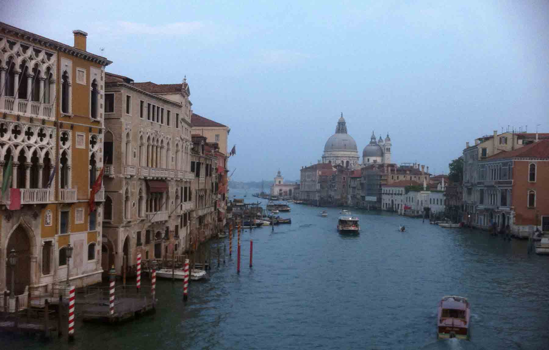
From Venice, I crossed to the mainland to meet a colleague before driving two hours inland, first west across the flat lands of Veneto province, then into the increasingly mountainous terrain of Trentino and finally to the dramatic landscapes of the Dolomites. The last four or five kilometres were on a gravel track that brought us, eventually, to Lago di Toval, set in a beautiful location, 1178 metres above sea level, amidst wooded slopes, with rocky alpine peaks visible all around us.

lago_di_toval

Lago di Toval, Trentino Province, Italy, September 2014

We were here for a paper-writing workshop at a small limnological research station beside the lake, eating and sleeping at a small albergo a few hundred metres away.   Confusingly, for a hotel situated beside a perfectly blue lake, its name was “Albergo Lago Rosso” but there was a story behind this name as my Italian colleague, Marco Cantonati, later explained.

The name of the albergo becomes clear when you see photographs of the lake taken in the 1950s and early 1960s when the water in some parts was a bright red colour due to growths of a an alga called (at the time) Glenodinium sanguineum.   The species epithet comes from the Latin sanguis, meaning blood, an allusion to the red colour of the cells which lends the lake its distinctive colour.   This alga belongs to a group called the “dinoflagellates”, which we have not previously encountered on this blog. The red colour comes from the same pigment that we encountered in Haematococcus (see “An encounter with a green alga that is red”).

albergo_lago_rosso

Albergo Lago Rosso, overlooking Lago di Toval, September 2014

For a long time, this alga was thought to exist in two forms, one of which was red, the other green.   However, the latest evidence suggests that there are at least three different forms, sufficiently different from one another to be assigned to separate genera. Two of these are only ever green whilst the third gives the lake its distinctive red colour.   This latter form was placed in new genus named after the lake where it was found, Tovellia.

Whilst the lake was famous for the distinctive red colouration that Tovellia sanguinea gave to it during late summer, this phenomenon has not been observed since 1964.   The other dinoflagellate species are still abundant, but T. sanguinea is now very rare.

lago_di_rosso

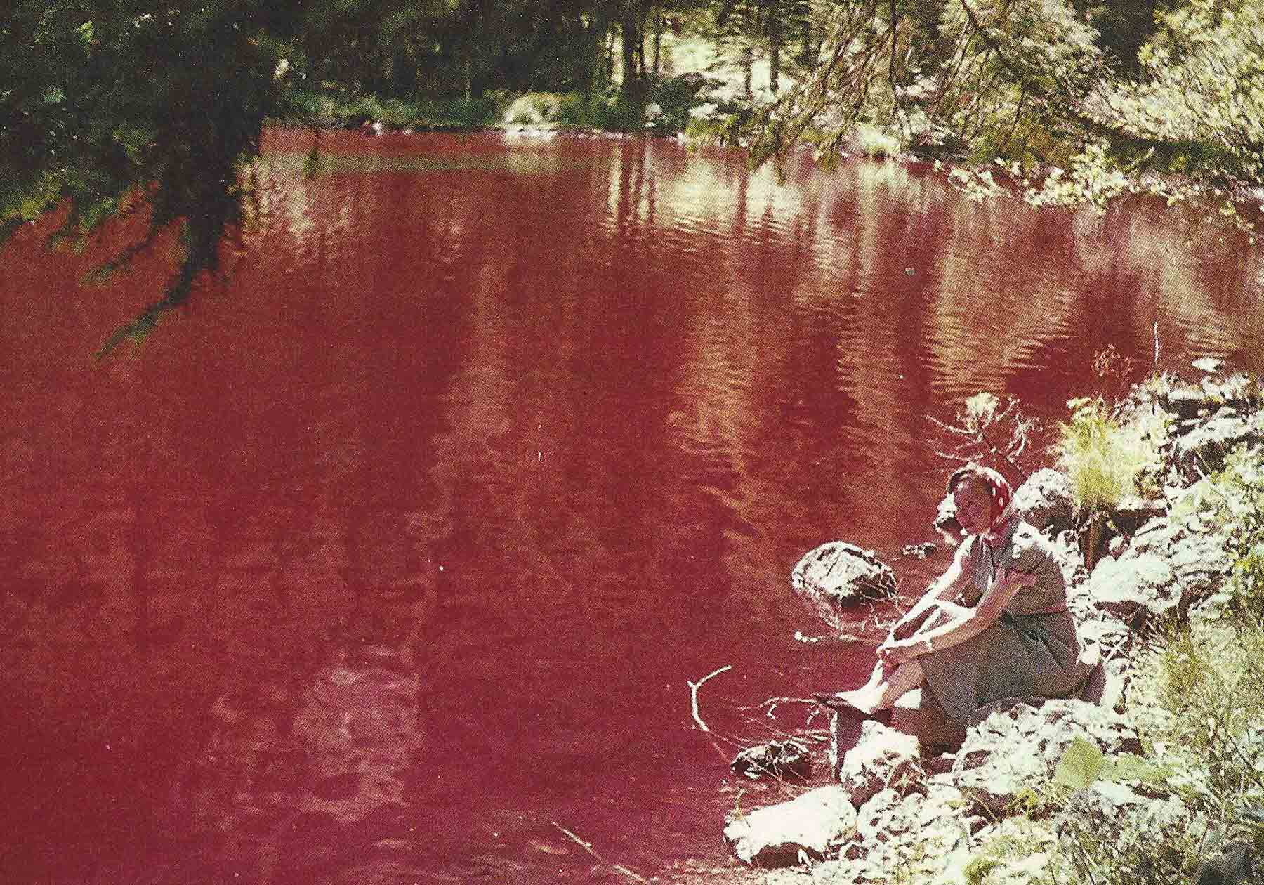
A postcard of Lago di Toval probably dating from the 1950s or early 1960s, showing the red coloration.

There is no definitive explanation for this change in lake colour but it is thought that changes in land use and, in particular, the way cattle were housed in the catchment, may have reduced the already small quantities of phosphorus entering the lake and tipped the scales in favour of the two green dinoflagellates rather than T. sanguinea.   There are other hypotheses and, as ever, it is difficult to untangle causation and correlation from the available evidence.   There is, however, also evidence that T. sanguinea was also rare before the 1860s, which does lend weight to the suggestion that the reddening of the lake was a response to human factors.   If this is the case, then Lago di Toval represents a relatively rare case of a lake that is returning to a more natural state.   That, of course, poses another fraught question: what exactly do we mean by “natural”? But that is a topic for another day.

References

Borghi, B., Borsato, A., Cantonati, M., Corradini, F. & Flaim, G. (2006). Il fenomeno del mancato arrossamento del Lago di Tovel alla luce dei risultati emersi dal Progetto SALTO. Studi trentini di scienze naturali – Acta biologica 81 Supplement 2: 471-472.

Cavalca, L., Ferrari, P. & Andreoni, V. (2001). Glenodinium sanguineum March. and the reddening phenomenon of Lake Tovel: biological and environmental aspects. Annals of Microbiology 51: 159-177.

Flaim, G., Moestrup, Ø, Hansen, G. & d’andrea, M. (2006).   Da Glenodinium a Tovellia.   Studi trentini di scienze naturali – Acta biologica 81 Supplement 2: 447-457.

Hansen, G., Daugbjerg, N., Flaim, G. & D’andrea, M. (2006). Studies on woloszynskioid dinoflagellates II: On Tovellia sanguinea sp. nov., the dinoflagellate responsible for the reddening of Lake Tovel, N. Italy. European Journal of Phycology 41: 47-65.

Venice’s green fringe

Back in April I commented on how little green I saw in Manhattan (see “A walk along the High Line”); however, I think that Venice probably beats Manhattan by some distance as the most built-upon city I have visited.   So close together are the buildings crammed onto the islands that comprise the city that there is very little space for any greenery or vegetation at all.   There is, moreover, nowhere equivalent to Central Park where locals can escape the urban fabric. I’ll go one step further and make the bold suggestion that Venice is the only city in the world where the most prolific type of vegetation is algae.

The algal growths are conspicuous at water level all around Venice: green lettuce-like fronds covering the steps, as in my picture below (taken close to Piazza San Marco).   I could recognise this as the genus Ulva, which we have encountered in other posts (see “The River Wear in Summer”) but not the species, though a quick hunt on the internet suggests that the most common species in the brackish water of the Venice Lagoon is Ulva rigida, a species that we do have in the UK.   I also found an estimate that a million tonnes of U. rigida biomass is produced each year in the Venice Lagoon, largely as a result of the nutrients that enter the lagoon.   Venice’s long tradition as a trading centre has resulted not just in the wonderful architecture and art that surrounded me throughout this trip but also in a densely-populated industrialised hinterland, the Veneto, as well as the city itself.   Much of the domestic and commercial waste made its way into the Venice Lagoon, leading to high concentrations of many pollutants.

Ulva_in_Venice

Ulva cf rigida coating steps near Ponte di Paglia, Venice, September 2014.

Phosphorus is an important limiting nutrient for algae, as I have explained in earlier posts, and I was interested to read that the city has banned the sale of phosphorus-containing detergents, in an effort to reduce concentrations washed down the drains and into the lagoon.   However, a final twist to the story is that there are also long-term plans to build a flood barrier across the entrance to the lagoon in an effort to reduce the number of occasions when the city is flooded.   Yet, when this is completed (expected in 2017) it will not only stop water from the Adriatic coming into the city, it will also inhibit the movement of polluted water from the lagoon into the open sea.

Reference

Cuomo, V. Peretti, A., Palomba, I, Verde, A. & Cuomo, A. (1995). Utilisation of Ulva rigida biomass in the Venice Lagoon (Italy): biotransformation into compost. Journal of Applied Phycology 7: 479-485.


Walking in Tintoretto’s shadow

The profusion of great art in Venice was an incentive not just to look but also to get out my own sketch pad and have a go.   There is no better way to appreciate Tinotretto’s very physical approach to the human body than to try to emulate his work.   The individuals in Tintoretto’s paintings are very muscular and are presented in dynamic poses, depicting images from the Bible, early Christian mythology or Venetian history.   Tintoretto makes great use of light and shade and, also, continues Titian’s experiments with colour.   Simply looking at images such as Cain and Abel in the Accademia reminds us that artists such as Tintoretto were as highly trained in anatomy as the doctors of the day. Indeed, artists could actually use their anatomical knowledge more productively than surgeons in an era when active interventions through operating were likely to lead to painful deaths from subsequent infections.

sketches_after_Tintoretto

Sketches based on Tintoretto’s paintings in the Accademia and Scuola Grande di San Rocco, Venice, September 2014.

Following the maze of small streets in Venice from the Accademia brought me to Scuola Grande di San Rocco, where walls and ceilings are covered by Tintoretto’s work.   It is hard to fully appreciate Tintoretto without developing a crick in the neck as some of his most technically sophisticated work consists of ceiling panels. Look up at the ceiling and you see an image of St Roch (“San Rocco”, much loved in Venice for his intercessions during a plague) in full single-point perspective. The ceiling is roughly ten metres above the ground yet the impression is that St Roch’s head is twelve metres above us and that there is open sky far above him.   Think about the steps needed to create this image: first, you work out the sketches on paper, then you have to scale up these working images and transfer the outlines to the ceiling.   When you were actually painting you would be crammed between the top of the scaffolding and the ceiling, constantly bent at uncomfortable angles and unable to step back, as most artists like to do, to check that the perspective that you worked out so carefully on paper, is working in practice. You will not see your work from a distance that allows you to check the perspective until the scaffolding has been removed.   And, as you travel around Venice, you see that Tintoretto painted many, many of these technically-challenging ceiling panels over the course of his career. His achievement is – and please permit me the use of a sorely-overworked adjective here – awesome.

Still reeling from the visual feasts in the Accademia and elsewhere in the city, I sat myself down on a step at the point where the Rio di Palazzeo joins the Canale Grande to make some of my own experiments in capturing human form.   A short distance along from here is the famous [bridge of sighs] so it is a popular route for the gondolas that carry tourists around the city.   Sitting here with a sketch book gave me the opportunity to observe a sequence of gondoliers as they swung their boats into the canal towards the Ponte dei Sospiri (Bridge of Sighs) and to capture their movements with a pencil.   Capturing the relative position of hands, arms and legs at same point in the stroke was not easy and, after the first few attempts, I realised how much I drew upon experience as much as on reality. I captured the basic form of the scene – relative positions of gondolier, boat and horizon – then filled in details with stock shapes that I had drawn before, underpinned by the very shaky knowledge of anatomy that a modern fine art graduate posseses.   Then I looked back and adjusted these to fit the reality that I saw in front of me.   This was all before I started to think about colour (surely a capital crime in Titian’s home town?).   The result probably says more about my lack of practice at figure drawing than it does about the verité of modern Venice but, after a day of admiring the energy that fills Tintoretto’s canvases, the awe that I experienced in the Accademia is tempered by plenty of humility.

sketches_from_Venice

Sketches of gondoliers, from close to Ponte di Paglia, Venice, September 2014.

sketch_of_gondolier

A watercolour sketch of a gondolier close to Ponte di Paglia, Venice, September 2014.

The Geography of Art

Venice_canal_grandel

Feeble excuse of the year coming up:

I have a meeting with some Italian colleagues next year but the flight times on Monday were not very convenient, so I decided to spend a weekend in Venice so that we can get down to work bright and early on Monday.   The reward for this noble act of self-sacrifice is the opportunity to gorge myself on art for the weekend.

As a part-time lecturer in a Geography Department, I find myself walking Venice’s narrow streets and pondering how art has geographies of its own. I have been fascinated by the history of art for some time, but my travels this year have made me acutely aware of just how time and space are tightly knitted together.   Why, for example, does Venice have relatively few paintings by Titian, its most famous Renaissance painter, yet so much by Tintoretto?   Why haven’t I yet seen a Canaletto here? Or, take the story back a few steps, what was it about Venice that allowed Titian to flourish in the first place?

Accademia_&_Titian

Left: A Canaletto-free zone: the Accademia gallery in Venice; right: Titian’s Tobias and the angel, photographed in the gallery.

I was at the National Gallery’s Making Colour exhibition a couple of weeks ago, which reminded me how the pigments from which artists derive their colours also have complicated stories, none more so than ultramarine, the intense blue pigment so conspicuous in Titian’s work. Once you know that it is derived from a rare mineral extracted from mines in Afghanistan it cannot be a coincidence that a city that built its wealth on trade with the Orient also bred artists who made bold experiments with colour.

The Titian / Tintoretto distinction is interesting.   I don’t have internet in my rented apartment as I write this so what follows is speculation but Titian was a superstar artist of his time who worked for many clients around Europe. Many of these paintings were sold over time and made their way to the art markets.   Napoleon, apparently, removed many Titians from Venice, which is why the Louvre has a good collection. Tintoretto, on the other hand, spent most of his career in Venice producing religious-themed art for institutions, several of which are still extant.   The Scuola Grande di San Rocco is the supreme example of this.   If you want to see Tintoretto, you have to travel to Venice.   Indeed, if you want to see Tintoretto, you have to travel around Venice.   There are cities where the great art is concentrated in a few galleries – London, Vienna, Paris, Berlin, New York, for example. And then there are cities such as Rome and Venice which are, themselves, the galleries.

Canaletto’s absence is easier to explain: he was the eighteenth century equivalent of the many, many purveyors of tourist gee jaws that line the streets around the tourist honey-pots.   His canvases are really glorified postcards, telling the world about a Grand Tour undertaken by wealthy young men in the eighteenth century. That is why there are so many scattered around stately homes in Britain.   I am, frankly, not missing them. There are so many other artistic delights in Venice – Carpaccio and Gentile Bellini have fascinated me as much as the Tintorettos – that I expect to be fully entertained without venturing beyond the end of the seventeenth century at all.

Welcome back to the art-science interface

My most recent visit to the River Ehen stimulated me to continue my explorations of what the subaquatic microbial worlds would look like in close up.   Before showing my latest creation, here’s a view of my working space which, as befits my positon in the no-man’s land between art and science, is neither a “laboratory” nor a “studio”.   My microscope is on the right of the desk, with a monitor for displaying specimens at the back. To the left is the bookcase where I keep my identification guides, within easy reach.   The desk has been cleared of clutter to make space for my watercolour pad, paints and pencils.   The main attraction of my study for painting is the good natural light, which floods through the south-facing window.

art_science_interface

My study / studio / laboratory.

I have usually worked out a rough design for a painting in a sketchbook before I have started, working both from direct observations down the microscope or photographs, with occasional dips into my books to check features or dimensions.   I then build up the picture gradually, starting with the foreground, and then slotting the other organisms into the spaces around the individuals at the front of the picture.   Working with watercolour and gouache means that I have bursts of activity, followed by pauses whilst a wash dries, then another burst.   Or, to put it another way, I get on with real work (the stuff I get paid for doing) and take occasional breaks to add the next piece of detail to my picture.   The one I have shown here has taken about five days from start to finish, with painting slotted around other activities.

The finished picture is below.   I’ve written much about the River Ehen over the past eighteen months or so and have been constantly surprised at how much variation we have seen in the algal communities over that time.   Each time I visit, I pick up a stone covered in green algae from the stream bed but when I look at these apparently identical growths under the microscope, I se that several different species have appeared and disappeared over the course of the study. Back in winter 2013, these communities were dominated by Spirogyra (see “The River Ehen in February”); other times of the year, the most abundant alga has been Bulbochaete (see “The River Ehen in August”).   Just a few kilometres downstream, we saw completely different algae again (see “At last a red alga that really is red …”). The structure of the communities when green algae predominated has, however, always been similar: the green filaments form a distinct layer over the top of a diatom-dominated understory.   This is what I have tried to capture in this picture. I have also added some filaments of the cyanobacterium Calothrix sp (see “Looking is not the same as seeing …”).

Ehen_140903

The River Ehen in September 2014.   The biofilm on stones has an understory of diatoms including Tabellaria flocculosa and Fragilaria tenera (right foreground) below a “canopy” of Bulbochaete.   Other algae living in the biofilm include the cyanobacterium Calothrix (left foreground).   Both Calothrix and Bulbochaete have long hairs, which release enzymes to capture phosphorus bound to organic particles suspended in the water. The Bulbochaete filaments are about 10-15 micrometres (1/100th – `/67th of a millimetre) across.

The presence of both Bulbochaete and Calothrix at the same location tells an interesting story. If you look back through my earlier posts, you’ll see that the long hairs characteristic of Bulbochaete and some other algae we’ve seen in the Ehen are thought to be adaptations that allow these species to thrive in situations where phosphorus is very scarce. But Calothrix also has the capacity to fix nitrogen, which is a useful adaptation when nitrogen is the scarcest nutrient. We know that nutrient levels in the River Ehen are low but the presence of both types of adaptation simultaneously suggests the organisms may be subject to both phosphorus and nitrogen limitation, perhaps reflecting short term oscillations in the relative availability of each over the year.

We’ve now got heaps of data telling us what species of algae are present, and how much algae there are over the courseof a year.   What we have done is akin to lifting up the bonnet of a car and naming all the parts of the engine. Our next task is to try to work out what all these different parts are doing (interpreting the role of hairs is part of this) and then, more importantly, to work out why the River Ehen’s engine doesn’t seem to be running as smoothly as we think it should.   I’ll come back to this in a future post.

Why Scottish Independence is bad news for English rivers

The environment has been conspicuous by its absence from the frenzy of debate and speculation in the run up to the Scottish independence referendum on Thursday.   That’s easy to explain: responsibility for the environment has already been devolved so there should be no additional implications from a “Yes” vote in the referendum. I’m not, however, fully convinced that there will be no implications north or south of the border in the event of Scottish independence.   Here’s my reasoning:

Although responsibility for the environment has already been devolved to Scottish, Welsh and Northern Irish administrations, the UK still presents a united front in Brussels, and a lot of co-ordination takes place behind the scenes to ensure consistency of policy.   There is a measure of pragmatism here: the politicians might think that the environment is a nice, neat package that can be managed equally well from London or Edinburgh but we traditionally use rivers as borders.   If you want to manage a river such as the Tweed, you really need the regulators on both the English and Scottish sides to agree on basic principles.   The UK and, indeed, many other federally-organised countries (Germany, for example) manage this.   The notable exception is Belgium where Flanders and Wallonia send separate delegates to meetings.

If Scotland votes “Yes”, then the newly independent state will have to apply for membership of the EU; the politics of this are complicated but let us assume that, at worst, Scotland becomes an Accession State within a few years and continues to apply EU Environmental legislation. The situation between England and Scotland will then be akin to that between Northern Ireland and the Republic of Ireland, where there is a long tradition of healthy collaboration between environmental bodies on both sides of the border. The details of implementation, however, differ between UK and Ireland and it is possible that a river deemed to be of an acceptable standard in Northern Ireland may feed into a river in the Republic, subject to more stringent standards (I know, for example, that standards for acceptable nutrient concentrations are tighter in the Republic than in the UK).   However, as there are relatively few catchments straddling boundaries between UK and neighbouring states (including Scotland, for the sake of this post), few diplomatic feathers will be ruffled as a result.

My nagging fear concerns the rump UK, rather than Scotland.   Staff working for environmental agencies tend to be either pragmatists, schooled in the “art of the possible”, or idealists.   Most of the ecologists I deal with in the UK’s agencies probably fall into the latter category, though I know that there are many non-ecologists involved in regulation who question the high standards that these ecologists call for.   And, indeed, the pragmatists have a great track record of reducing pollution levels in our rivers from previously high values. The problem is that the lower levels of pollutants that are now common in our rivers are still often too high to permit healthy rivers to thrive.   The problems are widespread but are most acute in lowland England where, in my experience, much of the water industry is sceptical about whether more investment (and associated price rises) will yield tangible benefits. Because Scotland has so much relatively high quality habitat to manage, and tourist economies that thrive upon this, there is less of a mismatch between the pragmatist and idealist viewpoints.   It is still there, believe me, but it is not quite so pronounced.   So now we extract the Scottish influence from the UK co-ordination meetings and, I fear, the balance of power will shift just a little way further away from a vision of babbling brooks supporting healthy ecosystems and towards grudging compliance but no more.

A very happy pearl mussel

I could not resist including this photograph of a pearl mussel grinning at my camera, partly because it reminded me of a paper I read a few months ago on the problems of conserving species such as pearl mussels which are not “charismatic”.   Though pearl mussels lack characters, such as large eyes that make it easier for humans to empathise with them, there is, nonetheless, a layer of language used by conservation professionals which sits over the usual objective language of science when discussing the plight of mussels.   This, the authors argue, helps us to think of species in human terms, even though they are “rhetorically challenged”.   They point to one officer in a conservation body who referred to pearl mussels as “poor souls” who needed our help – the classic language of charity. And, indeed, there must be something about pearl mussels that raise them so much higher on the conservation agenda than most other invertebrates and way much higher than the lower plants which interest me the most. And, looking at this fellow leering at me from the bed of the River Ehen, I could see that they maybe had a point. Who would not want to conserve such an anthropomorphic little mollusc?

pearl_mussel_in_Ehen

A happy pearl mussel in the River Ehen

Reference

Carrithers, M., Bracken, L.J. & Emery, S. (2011). Can a species be a person? A trope and its entanglements in the Anthropocene Era. Current Anthropololgy 52: 61-685

Pearl mussels with some unexpected bedfellows …

I was thinking about my recent encounters with epiphytic algae (see “Cladophora and friends“) during my most recent visit to the River Ehen, when I noticed algae attached to some of the pearl mussels on the stream bed.   We’ve seen this a few times before but today I had my Olympus TG2 camera at the ready and was able to take a few photographs.   There have been occasions when the quantity of algae on the shells has been greater than this, and there has been concern about how this algae may affect the mussel’s ability to feed.   It is one of several possible stresses on the pearl mussel population that we are investigating at the moment.

When I put a small piece of algae that I removed from the mussel shell onto a slide and had a look at it through my microscope, I saw that it was Oedogonium, which we have already met in some posts from earlier this year (see “More about Oedogonium” and links therein).   My guess is that this is not a sophisticated relationship between host and alga, but simply that the mussel shells represent a convenient and relatively stable substratum for opportunistic filamentous algae, which seem to thrive in the River Ehen at this time of year.

Oedogonium_on_pearl_mussels

Pearl mussels (Margaritifera margaritifera) on the bed of the River Ehen, several with bright-green growths of Oedogonium attached to the shells.   Note, too, the moss Fontinalis antiypretica in the upper part of the picture.

The description of an alga growing on an animal shell makes a nice counterpoint to the recent posts on algae growing on other algae.   There are other accounts of epizoic algae, though nowhere near as many as for epiphytic algae. This might simply mean that we have not looked in the right places.   The most spectacular of the few records that I do know about comes from my Belgian friend, Luc Denys, who scraped the backs of beached whales and found two previously unknown genera of diatoms that seem to grow exclusively on this rather unusual habitat.

Oedogonium_Ehen_Sept14

Epizoic Oedogonium from the River Ehen, September 2014 at low (x100, left) and medium (x400, right) magnification. Scale bar: 10 micrometres (1/100th of a millimetre).

Reference

Denys, L. (1997). Morphology and taxonomy of epizoic diatoms (Epiphalaina and Tursiocola) on a sperm whale (Physeter macrocephalus) stranded on the coast of Belgium.   Diatom Research 12: 1-18.

Nitzschia and a friend …

I drove from Bogle Bridge, my sample site on Stockerley Burn, up a low hill and down the other side into the next valley where another tributary stream of the River Browney flowed. I scrambled down to the stream and hunted amongst the stones for any algae. There were fewer green filaments here but I did find a small area of a stone covered with a dark brown mat that intrigued me.   My initial suspicion was that it was a mat of the filamentous blue-green alga Phormidium, but it turned out to be composed almost entirely of Nitzschia sigmoidea, a diatom that we last encountered in the brackish waters around Jarrow (see “All things bright and beautiful?”).

Nitzschia_sigmoidea

Nitzschia sigmoidea from Smallhope Burn, Knitsley Bridge, August 2014.   The upper image shows a single cell, the lower image two cells shortly after division.   Scale bar: 20 micrometres (= 1/50th of a millimetre).

If you look back at the post on Nitzschia sigmoidea at Jarrow, you’ll see that it was smothered in epiphytes.   Some of the cells here at Smallhope Burn also had attached epiphytes, though this time it belonged to a different genus.

I have called this epiphyte “Synedrella subconstricta”, though it has had several different names during the course of my career.   When I started, it was Synedra parasitica var. subconstricta but it has also been moved to the genus Fragilaria and, from there, to Pseudostaurosira.   Some have regarded it as a variety, others as a distinct species.   What is generally agreed is that it is usually found attached to Nitzschia sigmoidea, and occasionally to other large motile diatoms.   Frank Round regarded this as a major reason for creating a new genus, Synedrella, for this and a related taxon, S. parasitica.   Don’t let this species name misguide you: there is no evidence that it is a parasite; it is more likely to be a commensal relationship of some kind.   I am less convinced by more recent efforts to move it to the genus Pseudostaurosira, which generally forms long chains rather than sharing the habit of single cells with a short but flexible stalk.   This move reflects the tendency of my fellow diatomists to spend too long staring at the empty cells of diatoms and not enough time watching them in their live state. I have hunted around to see if there is any molecular evidence to support any of these genus names but, so far, have not had any luck.   How, I wonder, would we get the pure culture that we need as our source of DNA when the diatom seems to be an obligate epiphyte?

And, one more question that puzzles me: what advantage does a sigmoid outline confer onto a diatom?   You’ll see from the second of my two images that there is also a cell of another sigmoid genus, Gyrosigma, in the same habit. This is a relative of Pleurosigma, which we at Whitburn last year (“Microscopic monsters in mud”) and there are also sigmoid species in the genus Stenopterobia.   The sigmoid habit must have evolved several times though it is interesting that all of these are motile genera and two (Nitzschia and Stenopterobia) also include straight species.  Any ideas?

N_sigmoidea_S_parasitica

Nitzschia sigmoidea with an epiphytic cell of Synedrella subconstricta and, just above them, a cell of Gyrosigma (probably G. acuminatum).   Scale bar: 10 micrometres (1/100th of a millimetre).

References

Medlin, L., Jung, I., Bahulikar, R., Mendgen, K., Kroth, P., Kooistra, W.H.C.F. (2008). Evolution of the diatoms. VI. Assessment of the new genera in the araphids using molecular data.   Nova Hedwigia Beiheft 133: 81-100.

Medlin, L., Yang, I. & Sato, S. (2012). Evolution of the diatoms. VII. Four gene phylogeny assesses the validity of selected araphid genera.   Nova Hedwigia Beiheft 141: 505-514.

Round, F.E. & Maidana, N.I. (2001). Two problematic freshwater araphid taxa re-classified in new genera. Diatom Research 17: 21-28.

Cladophora and friends

I was back at Stockerley Burn last week (see “A case of mistaken identity?”) and was surprised to find that the Oedogonium that was so prolific back in June has now almost completely disappeared and the stream bed is now dominated by Cladophora glomerata. How do I know?   Look at the illustration below: even with the relatively weak magnification offered by a hand lens you can see the characteristic branched structure that distinguishes it from an unbranched species such as Oegogonium.   There is almost nothing else that could be confused with Cladophora in our freshwaters, so long as you perform this simple test.

I saw Cladophora glomerata at almost all of the sites I visited during this trip, which was not a great surprise as it is a very common species in enriched lowland rivers and streams.   However, the Cladophora filaments did not have their characteristic green colour at many of these sites, mostly tending to a brownish hue due to the large numbers of diatoms which were growing on and around these filaments.

Cladophora_filaments

Left: Filaments of Cladophora glomerata on my fingertip, showing the characteristic branched structure; right: a wet mount of Cladophora photographed with a macro lens. The entire structure is about four millimetres long.

Two of these diatom-smothered filaments are illustrated below.   You can see the upright, curved cells of Rhoicosphenia abbreviata, each attached to the filament by a small mucilage pad, along with some lower-profile cells, with an oval outline. These belong to the genus Cocconeis and I think I could see at least two species (C. pediculus and C. euglypta) though it is hard to be sure when examining them in the live state.   The cells on the lower of the two images do look more like C. pediculus to me.   There are also a few cells of Fragilaria, again standing erect on the filament.

The second image shows another filament from the same location but this one is covered almost entirely with Cocconeis cells (mostly C. pediculus, I think). The lower, more streamlined, profile of these makes them more suited to situations where they are more exposed to the current.   Indeed, I suspect that there are subtle differences in the composition of this epiphyte flora throughout the tufts of filaments though it is hard to do more speculate when these differences are at a scale that is difficult to either see or handle whilst standing in a stream.

What effect do all these epiphytes have on the growing Cladophora?   The diatoms are trapping sunlight that would otherwise have been used by the Cladophora so the net effect of all of these is akin to moving a houseplant from a well-lit window ledge to a dark corner.   On the other hand, Cladophora grows very quickly so an probably stay one step ahead of the epiphytes most of the time. There is evidence that prolific epiphyte growth leads to the eventual death of some aquatic plant.   This has been suggested in the Norfolk Broads where gradual increases in nutrients favoured epiphytes which, in turn, smothered their host plants (slower growing than Cladophora), leading to the long-term deterioration of the unique habitats of the Broads.

Cladophora_epiphytes_#1

Epiphytes on a Cladophora filament from Stockerley Burn, August 2014; a. Rhoicosphenia abbreviata; b. Cocconeis sp (from above: valve view); c. Cocconeis sp (from the side: girdle view); d. Fragilaria sp. Scale bar: 10 micrometres (1/100th of a millimetre).

Cladophora_epiphytes_#2

Cocconeis cells epiphytic on a filament of Cladophora glomerata.   Scale bar: 10 micrometres (1/100th of a millimetre).