Who do you think you are?

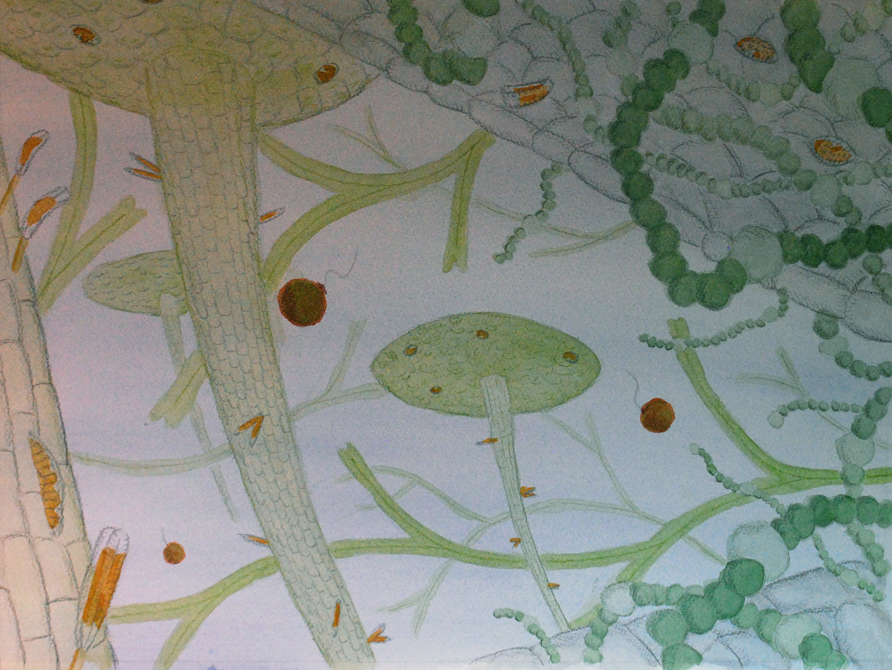
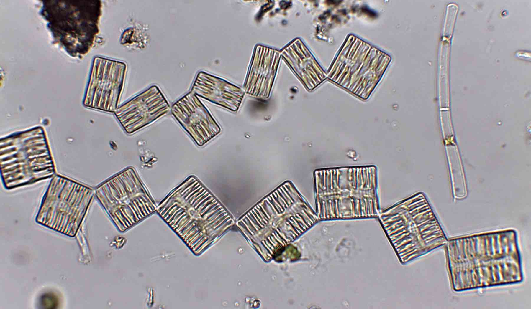
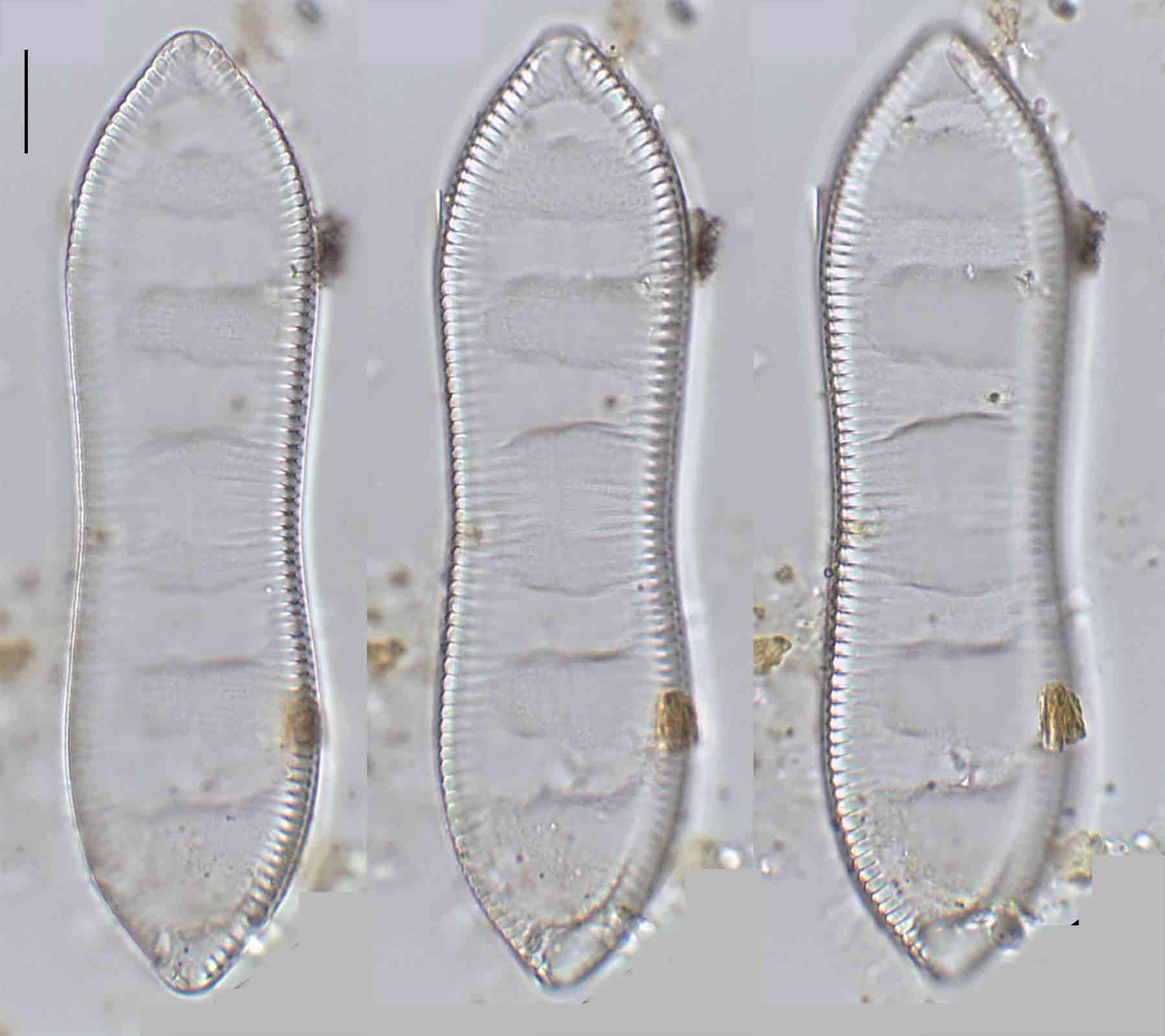
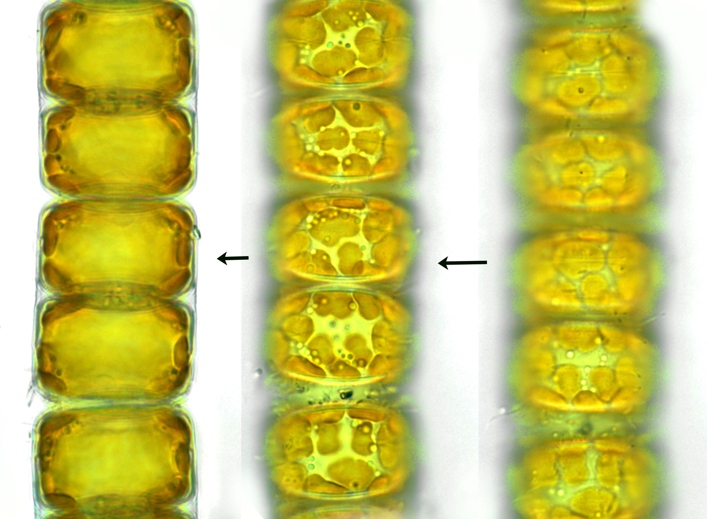
Few objects are more beautiful than the minute siliceous cases of the diatomaceae: were these created that they may be examined and admired under the higher powers of the microscope?  The beauty in this latter case, and in many others, is apparently wholly due to symmetry of growth.

Charles Darwin (1859) On the Origin of Species

Over the past couple of years I’ve written several posts giving overviews of the major groups of freshwater algae (most recently “Origin story …” and “Unlikely bedfellows …”).  I know that the of freshwater algae can be very confusing for outsiders, and I thought that sketching out some “family trees” might help folk work out how all these disparate groups link together.   The one group that seem to have slipped through the net, however, are the diatoms, the group which is the main focus of my professional life.   

That might be because I’m too close to the animated discussions that sometimes erupt around diatom systematics.   Those discussions, in turn, point to an absence of unambiguous evidence concerning the origins of the major groups.  David Williams and Pat Kociolek wrote in 2007 that “… a natural classification is still a long way off” and that statement still holds in 2022.  As a result, what follows is a “work in progress” that should, at least, help ecologists see how the many different types of diatoms they encounter fit into a bigger picture. 

Stephanodiscus – a centric diatom belonging to the Mediophyceae.   The photo at the top of the post shows species of Navicula – representatives of the Bacillariophyceae.  All photos: Chris Carter.

Broadly speaking, classifications of diatoms can be divided into three phases: 

  • the era when classifications were based what you could see with a light microscopy;
  • the era when classifications were based on what you could see with light and electron microscopy; and,
  • the era when classifications were informed by molecular genetics, as well as by light and electron microscopy.

Whilst the first era extended from the invention of microscopes in the 17th century to the late sixties, the second and third eras have both been compressed into the last half century, the “early adopters” of molecular approaches rubbing up against conservative-minded scientists still largely reliant on light and electron microscopy.

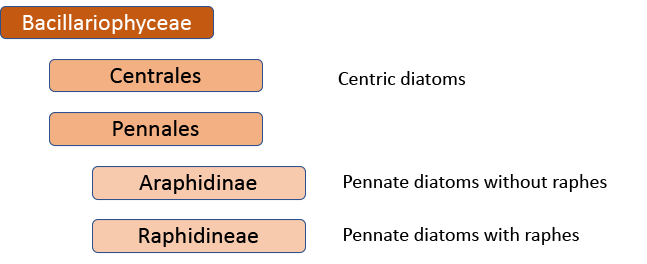
When I started, the standard work was Fredrich Hustedt’s 1930 Süsswasserflora von Mitteleuropa, which treated diatoms as a class, divided into two orders, the centrics (diatoms with at least one plane of radial symmetry) and the pennates (diatoms with at least one plane of longitudinal symmetry).   The pennate diatoms were, in turn, divided into those with and without raphes (the Araphidinae and Raphidineae respectively).  Hustedt was not the originator of this classification, but his works informed so much taxonomy in the middle and later twentieth centuries, that it is his name that is most associated with it.   Indeed, it was still the prevalent classification when Krammer and Lange-Bertalot started their revision of the Süsswasserflora in 1986.  

The classification of diatoms following Hustedt (1930), recognising diatoms as a class, and centric and pennate diatoms as two distinct orders. 

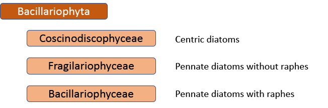
In 1990, however, Frank Round, Dick Crawford and David Mann produced a new classification, drawing much more on information from electron microscopy than Hustedt was able to do.   The end result of this classification is much the same as before – with centrics, araphids and raphids separated – but the two groups of pennate diatoms were now of equivalent rank to the centric diatoms.  Note, too, that diatoms as a whole have been promoted to a division rather than a class.   This juggling of taxonomic ranks might seem like an unnecessary complication for end-users, but it is absolutely necessary if the end-result is a “natural classification”, as referred to above.   Putting raphids and araphids as subgroups within a single pennate group implies that all pennate diatoms shared the same ancestor, regardless of whether or not they have a raphe. The Round/Crawford/Mann classification, by contrast, suggests that raphid and araphid pennates might be two separate lineages without a common ancestor.     

The classification of diatoms following Round et al. (1990), recognising diatoms as a Division and centrics, araphid pennate and raphid pennates as three distinct classes.

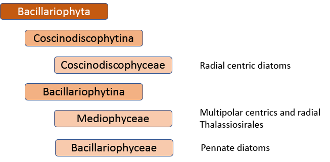
The first big impact of molecular genetics, however, was on the centric diatoms, hitherto thought of as a natural group.   Linda Medlin and Irena Kaczmarska broke the diatoms into two subphyla, with the centric diatoms now divided between these.   The Coscinodicophytina includes common freshwater diatoms such as Melosira varians (see “Some like it hot …”) whilst other common freshwater centrics such as Cyclotella and Stephanodiscus are in the class Mediophyceae within the Bacillariophytina, implying a closer relationship to pennate diatoms than hitherto suspected.   The Mediophyceae, whilst common in freshwaters, tend to be planktonic rather than benthic, so I have never really given them the attention that they deserve in this blog. The pennates, meanwhile, are back as a single group, the Bacillariophyceae, with both araphids and raphids combined.

The classification of diatoms following Medlin and Kaczmaraska (2004), with centric diatoms split between two classes.

That’s almost the end of the story, insofar as the Medlin/Kaczmarska approach is used by AlgaeBase, the website that summarises taxonomic knowledge of the algae.   However, further work has shown that this is still not a “natural” classification.  That would mean yet another shake-up, probably with at least nine separate, albeit “natural”, groups (see Theriot et al. in reference list).  There is now a very clear tension between those who believe that this is the best way forward in the long term and those who believe that minimizing the overall disruption experienced by end-users of taxonomic processes is also a consideration.   

Melosira varians – a representative of the Coscinodiscophyceae.   Cells are typically about 20 micrometres (= 1/50th of a millimetre) in diameter.  Photos: Chris Carter.

Interestingly, that final point may, itself, become the reason why we need a natural classification in the future.  At present (and at risk of upsetting systematists), higher classification provides a useful indexing system that means I can pluck a book off my shelves, and flick through to find the appropriate pages, when I am trying to identify a diatom.   All the older systems work just fine at that level.   The problem comes when we are using metabarcoding to identify diatoms.   Because our barcode reference libraries are far from complete, we cannot assign every sequence in a sample to an appropriate species.  In such cases, it would be good to assign it to a higher taxon and, for this to work, our reference libraries need to be arranged around a meaningful hierarchy, and systems that are adequate for a pragmatic light microscopist will cause problems for the logical decision-making processes that are used in bioinformatics routines.   

I’ve long argued that the “craft” of identifying diatoms is different to the science of taxonomy.  I could go further: taxonomists have, in recent years, tended to be more concerned about cataloguing variation within species and genera (discovering a lot more diversity than hitherto expected in the process) than with understanding the interrelationships amongst these groups.   However, we may now be closing the circle: “craft” of identifying diatoms got along fine with just occasional nods to systematics for a long time.  The brave new world of metabarcoding might mean that systematics is, once again, recognised as the important discipline that it should be. 

References 

Adl., S., Bass, D., Lane, C.E., Lukeš, J. et al. (2018).  Revisions to the classification, nomenclature, and diversity of eukaryotes.  Journal of Eukaryotic Microbiology 66: 4-119.   

https://doi.org/10.1111/jeu.12691

Medlin, L.K. & Kaczmarska, I. (2004).   Evolution of the diatoms: V. morphological and cytological support for the major clades and a taxonomic revision.  Phycologia 43: 245-270.

Round, F.E., Crawford, R.M. & Mann, D.G. (1990).  The Diatoms: Biology and Morphology of the Genera.  Cambridge University Press, Cambridge.

Theriot, E.C., Ashworth, M.P., Nakov, T., Ruck, E. & Jansen, R.K. (2015).  Dissecting signal and noise in diatom chloroplast protein encoding genes with phylogenetic information processing. Molecular Phylogenetics and Evolution 89: 28-36.

Williams, D.M. & Kociolek, J.P. (2007). Pursuit of a natural classification of diatoms: history, monophyly and the rejection of paraphyletic taxa.   European Journal of Phycology 42: 313-319.  

Some other highlights from this week:

Wrote this whilst listening to: Wet Leg and caroline’s eponymous debut albums plus Father John Misty’s Chloë and the Next 20th Century.

Currently reading:   Colm Tóibín’s The Magician, a novel about the life of Thomas Mann

Cultural highlight: first visit to the threatre since the start of the pandemic.  Went to see Red Ellen – about the life of Labour politician Ellen Wilkinson – at Newcastle Playhouse.  

Culinary highlight: “MSC carbonara”.  Recipe: buy seafood which has a blue Marine Stewardship Council logo on it, and make a carbonara from it.  This one was made from prawns, and enhanced by a pinch of chilli flakes and some lemon juice.  The next one will be made from something else.

Eyes wide open in Cassop’s muddy fringes …

The previous post mused on the distinction between “what I see” and “what I look for”.  They seem like two ways of saying the same thing, but actually hint at an awkward truth: ecology fieldwork is not always the objective process we like to pretend that it is.  My most recent trips to Cassop Pond have pushed this idea: for eight months I looked for algae amongst the Riccia fluitans flocs, on the rush stems and on duckweed.  There are other habitats that I could explore, but I chose not to.  What I looked for shaped what I saw.   In September and October I have redressed this by looking at a habitat relatively unfamiliar to me: the surface of the mud in the shallows at the edge of the pond.

If I used a pipette to hoover up some of the brown oozy mud and squirted that onto a microscope slide, I would probably see lots of fine sediment particles, plenty of dead and decaying organic matter but very few algae.   We know that they are there, and that they are often abundant, but they are hidden amongst all this other crud.   Instead, microscopists have devised two ways of separating the algae from the mud and I tried both of these on some of the Cassop mud which I scooped up with a plastic spoon and popped into a plastic bottle

The first of these involved letting the mud settle in the bottle, then pouring off as much of the overlying water as possible.  I then poured some of the mud that remained into a Petri dish and carefully lowered a coverslip onto the mud surface.   I left this overnight hoping that those algae capable of movement would glide up to the surface of the mud until they encountered the coverslip.   In the morning, with Andrew Marr grilling Rishi Sunak about the impending budget in the background, I carefully lifted the coverslip from the mud with a pair of forceps and dropped it onto a drop of water on a microscope slide.  

At the same time as I removed the cover slip, I placed a piece of lens tissue gently onto the mud surface and left it there for the rest of the day.  In the evening, I lifted it up and cut out a 5 x 5 mm square from the centre and placed this in a drop of water on a slide.  I then teased the fibres apart with a pair of dissecting needles before lowering a cover slip on top.   The idea behind both the coverslip and lens tissue methods is that muddy surfaces favour motile algae and that these will move up from the mud and onto the coverslip or tissue. 

Methods for sampling algae from fine sediments: a. coverslip placed on mud surface; b. lens tissue freshly placed on mud surface; c. lens tissue after lying on mud surface overnight; d. fragment of lens tissue (approx. 5 x 5 mm) placed in a drop of water on a slide then teased out with dissecting needles.

Both approaches yielded similar species, with a motile Navicula (possibly N. trivialis) most abundant, along with a small Trachelomonas (see “Microscopic mysteries in Cassop Pond).  Second most abundant by number of cells was Cymatopleura solea although the larger size of the cells, relative to those of N. cf. trivialis, might well mean that these contributed more to the total algal biomass.  Cells of Cymatopleura solea are shaped a little like the body of a violin when seen from above, and have a raphe around the margins that enables them to glide around.   Occasionally, whilst watching them move, one would slip onto its side revealing undulations on the surface that are not easily seen when looking from above.  I tried to describe the shape of C. solea to a class some years ago and, as I was grasping for appropriate adjectives to describe these multidimensional undulations, one student suggested “voluptuous”.  I have found C. solea in other samples from Cassop this year, but not in such numbers as from the mud surface.

Algae from the mud surface in Cassop Pond, October 2021.   a, b, valve and girdle views of a cell of Navicula cf. trivialis; c., d. valve and girdle views of a smaller Navicula; e. girdle view of Pinnularia sp.; f. valve view of Neidium cf. ampliatum; g., h. Synura petersenii.   Scale bar: 10 micrometres (= 1/100thof a millimetre).  

Alongside these were smaller numbers of Pinnularia, Nitzschia and a large Neidium (possibly N. ampliatum).   Bustling around, too, were some colonies of Synura petersenii, a flagellated chrysophyte that we have not met before in this blog.   There were, in addition, a few filaments of Spirogyra which has no means of moving so which must have moved onto the lens tissue by another means, perhaps through capillary action.    That brings me back to my initial musing about the difference between “what I see” and “what I look for”.   Using coverslips and lens tissue means that I see a different assemblage of algae to those that I find using my regular collection methods.  But, at the same time, I’m seeing the edges of Cassop Pond through a different filter.   These techniques are used because the assumption is that the algae that live on fine muds need to be able to move in order to survive but we test that assumption using a method biased to those algae that can move.  Staurosirella lapponica is a diatom associated with soft sediments that is incapable of movement, so any records from a coverslip or lens tissue approach would be “by-catch” and not give a true indication of its contribution to primary productivity.   There’s no perfect way of looking at worlds that we cannot ordinarily see.  But knowing that there is no perfect way of seeing does, at least, encourage us to keep on looking.

Cymatopleura solea from surface mud in Cassop Pond, October 2021.   a., b. valve views; c.,d. girdle views; b. and c. are two views of the same cell which obligingly rolled over whilst I was watching.  Scale bar: 10 micrometres (= 1/100th of a millimetre).  

References

Eaton, J.W. & Moss, B. (1966).  The estimation of numbers and pigment content in epipelic algal populations.  Limnology and Oceanography 11: 584-595. 

Moss, B. & Round, F.E. (1967).  Observations on standing crops of epipelic and episamnic algal communities in Shear Water, Wilts.   British Phycological Bulletin 3: 241-248.  

Yang, H., Flower, R.J. & Battarbee, R.W. (2010).  An improved coverslip method for investigating epipelic diatoms.  European Journal of Phycology 45: 191-199. 

Some other highlights from this week:

Wrote this whilst listening to:    New albums from War on Drugs (“I Don’t Live Here Anymore”) and local boy Sam Fender (“Seventeen Going Under”)

Cultural highlights:  Pedro Almodóvar’s recent short film The Human Voice, starring Tilda Swinton.

Currently reading:  American Dirt by Jeanine Cummins.

Culinary highlight:  Homemade ribollita.  

Microscopic mysteries in Cassop Pond …

I’ve been following the composition of algae in and around flocs of Riccia fluitans all year and have now tried to follow-up my picture of the algae in Pond Politics, in which I showed the liverwort and duckweed with attendant diatoms from samples collected in April, with one showing the situation in late summer.   In the meantime, we have seen Cladophora glomerata come and go (see: “Cassop Pond in June”) followed by the arrival of Nostoc.  In my post Change of Tenants I found filaments apparently free-floating and very similar ones attached to the fronds of Riccia fluitans.   I had wondered if the free-floating filaments were Anabaena sp. but now think that they are more likely to be Nostoc, mostly living in and around the Riccia fronds but detached by the various manipulations required to get a sample of the Riccia floc from the pond, back home and then under my microscope.    

We can get a better idea of the relationship between these filaments and the liverwort from some photographs that Chris Carter took for me.   Previous encounters with Nostoc in this blog have been with terrestrial species that form well-defined firm colonies, readily visible with the naked eye (see “How to make an ecosystem (2)”).   This is different: the colonies are, at best, ill-defined and on a scale not easily seen without magnification.   Based on the descriptions in the Freshwater Algal Flora of the British Isles, I think that these belong to Nostoc coeruleum, but Brian Whitton (who wrote the account) urges against placing too much confidence on differentiations made on the basis of often vague descriptions in the literature.   The habitat notes for N. coeruleum do, however, match the conditions in Cassop Pond: “In standing fresh or slightly brackish water, among submerged plants, free-floating or loose on bottom sediments …” 

Nostoc cf coeruleum on fronds of Riccia fluitans from Cassop Pond, Co. Durham, July 2021.  Photographs: Chris Carter.

Based on these photographs, it looks like Nostoc forms a layer on the surface of the Riccia leaves, gaining proximity to light in return for sharing the nitrogen it fixes from the atmosphere.   Curiously, I did not see Nostoc growing amongst the Lemna fronds, which suggests some host-specificity in the relationship.  Whether it is correct to describe this as a “symbiosis” rather than a mutually-beneficial interaction is not clear and would take rather more work.   That I did not see Nostoc until the middle of the year suggests that this may just be an opportunistic association rather than a long-term relationship.   

My picture tries to capture some of what is happening in these flocs.  Because the flocs grow in and amongst Phragmites, the left hand side of the picture has the stem of Phragmites along with some diatoms attached (Gomphonema, Epithemia and Achnanthidium).  On the right-hand side, by comparison, there are fronds of Riccia fluitans and associated Nostoc coeruleum.   There are also some diatoms – Cocconeis and Rhoicosphenia – growing on the Riccia fronds.   Finally, there are some floating leaves of Lemna minor, each with a hanging root.  Once again, there are diatom epiphytes – Cocconeis on the underside of the leaves and Fragilaria on the roots.  Finally, I have included some flagellated cells of the euglenophyte Trachelomonas, which are frequent amongst the Riccia fronds at this time of year (see also: “Puzzling puddles on the Pennine Way“).  It is quite a “busy” image but still, with eleven species represented, it is a highly simplified version of what is actually present in the margins of Cassop Pond.  It is also a snapshot – a moment in time – whereas the real assemblages will be forever shifting with the vagaries of wind and local disturbance, as well as changing in response to the seasons. 

Some other highlights from this week:

Wrote this whilst listening to:    Lambchop’s 1996 album How I quit smoking.  

Cultural highlights:  The Green Knight, new film adaptation of the Arthurian legend Sir Garwain and the Green Knight.

Currently reading:  Between the Assassinations by Arvinda Adiga, a. novel about a fictional town in India in the 1980s. 

Culinary highlight:  The bar is set fairly high hereabouts and I usually forget the basics, such as fresh sourdough bread with jam made from fruit from the allotment.  Not by me, I hasten to add.   

Invisible worlds at Malham Tarn

Dicky Clymo’s first – or maybe second – ecology lecture to Westfield College undergraduates in the early 1980s used Malham Tarn as a case study in how ecosystems change in space and time.   Malham Tarn is one of just two natural lakes in the Yorkshire Dales (the other is Semer Water – see “Lake lakelake lake”).  The permeable limestone bedrock means that most surface water seeps away; however, Malham Tarn sits on the underlying impermeable Silurian slate and water which has drained through the limestone bubbles up in a series of springs to the north-west of the tarn, picking up calcium and other ions on the way to create hard water with a unique ecology (see “Everything is connected …”). 

Over the millenia, however, fine sediments deposited by the streams that bring the spring water to the tarn accumulated and the north-west corner of the lake became shallower to the point where it was no longer open water, but a shallow fen colonised by amphibious plants.   In parts, those colonising plants have been replaced by alder carr, allowing Dicky to demonstrate the principle of ecological succession.   However, as the north-west corner of the lake slowly turned into a mire, so the influence of the calcium-rich springs lessened whilst that of rainwater increased.   The water in parts of the mire is now extremely soft and the juxtaposition of very soft and very hard water creates a wonderfully wide range of habitats within which to search for aquatic organisms. 

John Lund, for example, estimated that “there is little doubt that a thousand [species of algae] could be found in this area” which made it an excellent location (following Windermere and Ennerdale Bridge) for the third of the British Phycological Society / Quekett Microscopical Club microscopy weekends.   This meeting was originally planned for spring 2019 but the pandemic intervened, and it is only now that it feels safe to be meeting face-to-face once again.   We spent part of the weekend looking at Malham’s famous tufa-forming streams and the other part looking at samples we collected from the softer waters Tarn Moss.   For Dave, in particular, these habitats have an irresistible pull, as rich sources of desmids, but there was a wide range of other algae present too.  

Collecting algae samples from the fen area of Tarn Moss during the BPS/QMC microscopy weekend at Malham Tarn Field Studies Centre, September 2021.  The photograph at the top of the post shows Malham Cove, 3 km to the south of Malham Tarn on a visit in 2019.

My guide for describing the algae that we found was an old paper by John Lund from the now defunct journal Field Studies.   He recorded 199 genera from Malham Tarn in this paper and it was interesting to see how our haul from Tarn Moss compared with his account.   For example, we found the cyanobacterium Anabaena to be quite common in squeezings from the pools on Tarn Moss whereas he only recorded it from plankton in the tarn itself (though it was quite abundant in some years.  His paper does not record how many visits he made or what time of year, so there may be good reasons for differences.  The section on permanent pools is very short and does not mention any algae at all by name.   Nonetheless, his records are a very useful starting point for a modern visitor to the area.   

As well as Anabaena, we also found colonies of the cyanobacterium Aphanothece microscopica in a squeezing of Utriculaira from one of the pools on Tarn Moss, as well as green algae, diatoms and dinoflagellates.  Of particular interest were small, writhing cells of Euglena mutabilis (last encountered in “A visit to Loughrigg Fell”).   This is an unusual Euglena because it does not possess a flagellum.  It is also tolerant of very acid conditions – being encountered when the pH is as low as 2.0.  Allan Pentecost made a special study of E. mutabilis from Tarn Moss, published in the journal Field Studies in 1982.  

The most abundant diatoms in the squeezings from Tarn Moss was Tabellaria flocculosa, although there were also a few chains of T. quadriseptata, distinguished by the short spines on the edge of the valve (easily visible under a light microscope but not clear in the photograph below).  More about these in The bluffer’s guide to Tabellaria …. Finally, a few dinoflagellates were observed.  Most were too fast-moving to be captured on camera, but I have photographed Cystodinium cornifax (Lund recorded this as Glenodinium, but from similar habitats to us).   The dinoflagellates are a group that have not been encountered much in this blog over the years and this is the first photograph of one from my own samplings.  That does not mean that they are rare, only that I do not look in the right places.

Algae (other than desmids) and a protozoan from Tarn Moss, Malham, September 2021. a. Anabaena sp. (lower filament with hormogonium); b. Ankistrodesmus cf. fusiformis; c. Stentor; d. Aphanothece microscopica; e. Pediastrum boryanum; f. Eudorina unicocca; g. Tabellaria flocculosa; h. Tabellaria quadriseptata; i.-k. Euglena mutabilis; l. Cystodinium cornifax.  Scale bar: approximately 50 micrometres (= 1/20th of a millimetre).

Then, of course, there were the desmids.  Squeezings from Sphagnum and Utricularia revealed rich hauls of these beautiful algae, and a few of the more common are illustrated in the figures below.   All are from Tarn Moss as most desmids prefer soft water.  However, John Lund also records Oocardium stratum, a filamentous desmid that grows in highly calcareous environments, from Gordale Beck.   We did not see it on our visit, though it has been recorded in the recent past.  

Desmids from Tarn Moss, Malham: a. Closterium ehrenbergii; b. C. acutum; c. C. kuetzingii; d. Micrasterias rotata; e. M. truncata var. bahusiensis; f. Staurastrum telierum; g. Euastrum denticulatum.  Scale bar: approximately 50 micrometres (= 1/20th of a millimetre). 

I did not have my usual microscope and camera with me on this weekend trip.  Consequently, all the images of algae in this post were taken with my iPhone via a HookUpz adapter.  Unfortunately, I did not have an eyepiece graticule on my microscope, so the scale bars are all estimates and need to be treated with caution.  

This was, I guess, another example of “scientific jazz” (see “Jammin’ in the key of algae …”) insofar as we had a theme (Malham Tarn and its environs) but no set objectives other than observing as much as possible of the algal diversity in a relatively short period of time.   Different people improvised in different ways – the desmids were popular, but one of our number used the “coverslip capture” method to look at the diatoms present on the fine sediments of Malham’s littoral zone, and others looked at the Cyanobacteria and diatoms associated with Goredale and Mastiles Becks.   The weekend was more about asking questions than finding answers.  And about drinking craft beer.  But that’s a story for another day …

More desmids from Tarn Moss, Malham, September 2021: a. Netrium digitus; b. Pleurotaenium trabacula; c. Tetmemorus brebissonii; d. Cosmarium reniforme.   Scale bar: approximately 50 micrometres (= 1/20th of a millimetre).  

References

Lund, J.W. (1961). The algae of the Malham Tarn district.  Field Studies 1: 85-119.

Pentecost, A. (1982).  The distribution of Euglena mutabilis in Sphagna, with reference to the Malham Tarn North Fen.  Field Studies 5: 591-606.

Pentecost, A. (1991).  A new and interesting site for the calcite-encrusted desmid Oocardium stratum Naeg. in the British Isles.  British Phycological Journal 26: 297-301.

Piggott, M.E. & Piggott, C.D. (1959). Stratigraphy and pollen analysis of Malham Tarn and Tarn Moss.  Field Studies 1: 84-101.

Some other highlights from this week:

Wrote this whilst listening to:    Nubiya Garcia’s Prom, available on the BBC iPlayer.

Cultural highlights:   Rewatched Educating Rita, which is as good now as it was when I saw it on first release.  And Arab Strap’s gig in Newcastle this week.

Currently reading:  The Secret Life of Bees by Sue Monk Kidd

Culinary highlight:  make-it-up-as-you-go-along gooseberry mayonnaise on top of smoked mackerel on top of a less-than-perfect minestrone soup converted into a risotto (the irony being that we followed Felicity Cloakes’ “How to cook perfect minestrone soup” recipe)

Cover versions …

Lots of good performances are raised by a cover of someone else’s hit, and Slime Time was no exception.  The cover version that we chose was a short film, “Moon, Mud and Microbes” by filmmaker Susi Arnott and University of Westminster scientists Jane Lewis and Dain Son, which shows the algae which live in intertidal mud in the Thames in central London.

It depicts the changes over a single tidal cycle using time-lapse photography to compress the entire cycle into six minutes.   The film opens with a view looking upstream towards Tate Modern as the moon sets over a darkened London and, as dawn breaks, the tide gradually recedes, gradually revealing, first, some old wooden pilings and then the unprepossessing brown tidal mudflats.   The location then shifts to St Saviour’s Dock, just east of Tower Bridge, where we zoom in on one section of the mudflat and the magic starts to happen: patches of green and a darker chocolate brown start to appear on the grey-brown mud.   These, we told our Slime Time audiences, are algae “commuters” who make a daily migration up through the fine sediments, in order to do their important “work” of pumping oxygen into the atmosphere.   Their upward movement is stimulated largely by light, but also by internal body clocks – something to which an audience of campers can relate: we invariably wake up as soon as daylight floods through the thin walls of our tent on the first morning under canvas but, as days go by, our bodies slowly adapt to override this stimulus.  

Two stills from Moon, Mud and Microbes showing the mud at St Saviour’s Dock, approximately 30 minutes apart in real time, showing the algae rising to the surface.   The montage at the top of the post shows the Omni Tent ten minutes before our performance, slowly filling as Rachel Williams, our compère, pulls in the punters, and finally co-presenter Thomaz Andrade gets Slime Time under way.   

The film then cuts from the mud of St Saviour’s Dock to a view of the organisms responsible for the change in colour under the microscope.   We can see large green cells of Euglena ehrenbergii and needle-shaped diatoms, Cylindrotheca gracilis with their yellow-brown chloroplasts.  Other diatoms appear in subsequent scenes, but these were the most abundant types.   The Cylindrotheca cells are about a tenth of a millimetre long, giving our audience a rough idea of the scale of what they were observing.  All these algae were moving around the screen, to the surprise of our audience who assume “plants” to be static and only “animals” to move (apart from the cheerful clever clogs in the second row who pointed out that Venus Fly Traps also move).   We didn’t want to get into the deep technical arguments about whether algae were, in fact, plants (see “Identity crisis”) or into the details of how they move.   Simply revealing an aspect of natural history hitherto hidden from our audience was enough.  

Euglena ehrenbergii and Cylindrotheca gracilis: the two most common algae in the microscopic samples shown in Moon, Mud and Microbes.  The Cylindrotheca cells are about 100 micrometres (= 1/10th of a millimetre) long.

From looking at specimens using a light microscope the film then moves on to show some stills taken using a Scanning Electron Microscope, allowing details of individual pores (no more than a micrometre – a millionth of a metre – across).   The image below shows one of the diatoms we saw: Hydrosera triquetra.  This is a species that was formerly regarded as a tropical species, but which was found in the Thames in 1971 and which is now common as a golden-brown zone on the lower part of river walls between Greenwich and Putney.   There are also a number of records from other north European estuaries.  

A cell of Hydrosera triquetra from the St Saviour’s Dock mud sample.  Note the smaller diatom growing as an epiphyte on the side of the cell.   Hydrosera triquetra cells are typically about a 10th of a millimetre in diameter.  A still from Moon, Mud and Microbes.  

For the last 90 seconds or so the film cuts back to views of the mud, showing the algae “commuters” gradually disappearing back into the mud before the incoming tide covers them all with water again.  The wooden pilings which we saw emerge at the start of the film now disappear back underwater and the light slowly fades at the end of the day.   The final shot is a view looking downstream from the South Bank near the National Theatre towards Blackfriar’s Bridge and St Paul’s Cathedral.   The tide is, again, high and the moon is rising.   And so the cycle begins again … 

Reference

Coste, M. & Ector, L. (2000).   Diatomées invasives exotiques ou rares en France: principales observations effectuées au cours des dernières décennies.   Systematics and Geography of Plants 70: 373-400.

Tittley, I. (2014).  Non-native marine algae in southeastern England. Bulletin of the Porcupine Marine Natural History Society 1: 28-32.

Some other highlights from this week:

Wrote this whilst listening to:    Soar, the collaboration between Welsh harpist Caitlan Finch and Senegalese kora player Seckou Keita.   The answer to the question: “what would Bach’s Goldberg Variations sound like if played on traditional West African instruments.   And Nubya Garcia’s first album 5ive.   Audio methadone to help me through post-Green Man cold turkey.   

Cultural highlights:  New BBC series Vigil which is, in essence, a Golden Era crime mystery but set on a nuclear submarine rather than in a country house.  

Currently reading:  The Sea is Not Made of Water: Life Between the Tides by Adam Nicholson.

Culinary highlight:  Lamb henry, a half shoulder of lamb slow cooked until it is meltingly tender and falling off the bone.  Served with a rich gravy and mash in the Shepherd’s Arms, Ennerdale Bridge, our regular fieldwork home-from-home.   Washed down with a pint of Loweswater Gold.  

Jammin’ in the key of algae …

It’s official: I have now performed at a major music festival.  In its 20-year history Green Man has seen countless guitar solos, plenty of saxophone and keyboard breaks and even, yea gods, a few drum solos.  On Friday and Saturday, for the first time, it experienced its first microscope solo.  In the spirit of improvised jazz, I went out on stage with a few themes in mind but from that point on what happened was down to what I saw through the microscope eyepiece (and which the audience saw on a screen behind me) and not to any pre-prepared script.  

Some context: I’ve been to the Green Man festival in the Brecon Beacons a few times in the past as a punter, and there is always a thriving science zone, Einstein’s Garden.  Last time I was here, in 2019, I noticed some unprepossessing flocs of algae floating in an artificial pond close to the impressive Mountain Stage (shown above) and thought: “they would look really good projected on to a big screen”.  And so the idea of “Slime Time” was born.   The second key component of Slime Time is a six-minute film of algae growing on exposed mudflats in the Thames in central London came when I met filmmaker Susi Arnott, but that will be the subject of a future post and I’ll focus on the improvised microscope solos in this one.

We pitched Slime Time to Green Man in early 2020 and they liked it enough to put us on the bill for the festival.   Shortly afterwards a certain pandemic kicked off and Green Man 2020 was cancelled, along with every other cultural event.   For a long while we thought that the 2021 festival wouldn’t go ahead either but, in mid-July, the Welsh Government finally gave the go-ahead and we all had about a month to get everything together ready for the gates opening ot the public on 19 August.  

The pond which had first inspired me to pitch the idea had, however, changed in the two years since I first saw it.  Gone were the floating flocs of algae and, in their place was a dense mat of duckweed.  Underneath this, however, I found some fine filaments, rough rather than slimy to the touch, which turned out to be blanket weed (Cladophora glomerata) when I got back to the tent and peered at it through my field microscope.  This is not the most exciting filamentous alga to show an audience but, fortunately, I had stuffed a small bottle of Spirogyra from Cassop Pond (see “Promising young algae …”) into my cool box just before leaving.

The artificial pond on the Green Man site, showing the dense cover of duckweed (photo: Sophie Perry)

A festival setlist, however, needs more than two tunes so I needed to do some more exploration around the Green Man site before hitting the stage.   First stop was the Monmouthshire and Brecon Canal which runs along the edge of the Usk Valley and is a ten minute walk from one of the festival’s gates.   That, however, was a disappointment.  The propellers from a steady stream of canal boats stir up the fine bottom sediments to create a turbid brown soup within which no respectable algae are likely to survive.   So, after a pleasant walk along the towpath, fuelled by some welsh cakes sold to me in aid of a cancer charity by two young girls and their grandmother, I headed back, still needing a banger to bring the show to a close.  

The Monmouthshire and Brecon Canal near the Green Man Festival site, August 2021.

The River Usk, which runs along the northern edge of the festival site, provided the inspiration for my closing number.   This is a river of high conservation value for several reasons, including the presence of the Twaite Shad (Alosa fallax).  Pete, the security guard watching the exit to the Green Man site as I left, told me that he had seen a large brown trout resting in a backwater close to the bridge, as well as spotting a salmon leaping.  As we chatted, there was the briefest of flashes of iridescent blue as a pair of kingfishers flew low along the water and my journey down to the river’s edge also disturbed a dipper.   These, I felt, were all good signs, reflecting Natural Resources Wales’ (NRW) assessment of the ecology here as “good status”.  

The River Usk looking upstream towards the entrance to the Glenusk estate, August 2021

Down at the water’s edge, however, the omens did not look so good, with stones smothered by a thick film of loosely-attached brown flocs which disintegrated to the touch.   Under the microscope these revealed themselves to be mostly made of the chain-forming diatom Melosira varians, along with Diatoma vulgaris(abundant in rehearsals with the field microscope, less common during my live set …) and a chain of Fragilaria(abundant during the live set, not apparent when photographing the diatoms on my return).   Overall, the impression that these algae gave me was that NRW’s overall assessment of “moderate status” is about right, reflecting concerns about the elevated concentrations of nutrients present in the river. To be fair to NRW, rivers never look at their best in late summer (see “Summertime Blues …”).   A combination of low flows, warm water and long hours of bright sunshine create ideal conditions for algae to grow and to outpace the invertebrates that feed upon them.  

As with a guitar solo, there comes a point when you pass from embellishing the main theme into self-indulgence and, with an audience composed largely of primary-aged children and their parents, I felt that my improvisations around the theme of the River Usk had reached a natural conclusion.   And, in the time-honoured festival fashion, it was time to say “that’s all we’ve got time for, you’ve been a great audience, enjoy the rest of the festival …” before throwing my microscope against an amplifier and leaving the stage to an overwhelming howl of feedback.    In my dreams …

The margin of the River Usk at Glenusk (beside the Green Man festival site) in August 2021 showing the thick film of Melosira-dominated biofilm smothering the rocks.   
River Usk diatoms: photographs taken on the same sample as I used for the live show, but stored in my tent for three days before I had a chance to examine it again. Consequently the chloroplasts of some diatoms are past their best.   a., b.: Ulnaria ulna; c. Diatoma vulgaris; d., e.: Didymosphenia geminata; f. Cymbella cf. neolanceolata; g. Achnanthidium minutissimum; h. Navicula tripunctata; i., j. Navicula sp (possibly N. cryptocephala, but N. gregaria was also present in the sample); k. Placoneissp., l. Cymatopleura solea; m. Melosira varians; n. Cocconeis pediculus growing on a filament ofOedogonium.   Scale bar: 20 micrometres (= 1/50th of a millimetre). We did not see Didymosphenia geminataCymbella neolanceolataCymatopleura solea or Oedogonium/Cocconeis pediculus during either of the live shows, so these are included here as “studio overdubs”.

Note: I was asked, after our show, for some guidance on how to buy a microscope.  If you are interested in exploring the microscopic world for yourself, this post, “Getting started with microscopy” offers some hints on buying your first microscope.

Some other highlights from this week:

Wrote this whilst listening to:    the Far Out Stage at Green Man which was about 200 metres from our tent as the crow flies.  The first draft of this post was written longhand into my notebook whilst Crack Cloud and Porridge Radio were on the stage on Sunday afternoon.  Other highlights of the weekend include Wet Leg (opening the festival on Thursday afternoon), Nubiya Garcia’s excellent jazz, Thundercat, Laura Marling’s LUMP, Richard Dawson, Big Joanie, interplay between harp and kora from Caitlan Finch and Seckou Keita,  and headline sets from Caribou and Mogwai (whose soundcheck provided a backdrop to our Saturday show).  The overwhelming memory of Green Man 2021 will be the joy on musician’s faces as they played to live audiences again; in many cases this was their first show since the start of the pandemic.

Cultural highlights:  Green Man (see above)

Currently reading:  Pie Fidelity by Pete Brown: a paean to British Food.   Bought at Book-ish, the excellent Crickhowell independent bookshop that operates a pop-up shop on the festival site.

Culinary highlight:  Green Man is also a street food and beer festival.   From the many stalls on offer, the highlight (by a small margin, because all were very good) was Taste Tibet, finalists in the streetfood category of the BBC Food Awards.   Of the beers on offer, Stouty McStout Face, from Mad Dog Brewery, Porthcawl, is worth the price for the name alone, but is also a very decent pint.   

Duckweed delights …

I’m hopping around in both time and space in recent posts.  The previous one was based on a visit to the Lake District in June whilst this one crosses the Pennines to report on a visit to Cassop Pond in May.   There is always a lag when studying diatoms, because of the preparation steps involved, but it has been longer than normal this time due to the pressure of other work.   In The Diatoms of Cassop Pond, I wrote that I had found 98 different species in samples collected in January, February and March.  Having looked at samples from April and May, this list has now been extended to 134 species but this is about more than just “twitching” because I’m also intrigued by how habitats around the lake margin differ in the diatoms they host.  

One of the most distinctive features of Cassop Pond are the extensive flocs of the liverwort Riccia natans, and I’ve noted the predilection of nitrogen-fixing diatoms (principally Epithemia adnata) for this substratum in previous posts (see “Working their passage …”).   One part of the margin, however, is dominated by duckweed (Lemna minor).   There is always some duckweed mixed in with the Riccia flocs, but it is usually a minor component.  How different, I wondered, were the diatoms when I looked at an almost pure growth of duckweed?  

Monoraphid diatoms from Cassop Pond, April and May 2021.   a., b. Cocconeis euglypta; c.,d. C. lineata; e. C. pseudolineata; f.g. Cocconeis euglypta/lineata/pseudolineata raphe valves; h.-l. Lemnicola hungarica (two raphe and three rapheless valves).   Scale bar: 10 micrometres (= 1/100th of a millimetre).

I had a hunch that I knew the answer, because I wrote about the epiphytes of duckweed in a previous post (“The green mantle of the standing pond …”), describing work in a Norfolk pond that had identified two species as being particularly characteristic epiphytes: Lemnicola hungarica and Sellaphora saugeressii (formerly S. seminulum).   I had found both of these in other samples from the pond, but they were more abundant on the Lemna than elsewhere.  L. hungarica accounted for five percent of the total number of valves, whilst S. saugeressii accounted for three percent.   By contrast, the Lemna samples had much less Epithemia (a single valve, compared to ten percent in the sample from Riccia) whilst the assemblages on the two plants were both dominated by Cocconeis spp.   The difference in representation of Epithemia is intriguing, based on what we know about its ecology, as it suggests that the Lemna assemblages are less nitrogen-limited than the assemblages on Riccia.   My hunch is that the most Lemna-rich area of the pond is a heavily-shaded inlet and, with the light needed for photosynthesis in such short supply, the plants grow more slowly and, as a result, the demand for nitrogen is lower.

One other species that I found for the first time in the May sample was Adlafia bryophila, a small diatom whose habitat is described as “locally frequent on intermittently wet bryophytes, aerophilous”.  In Cassop Pond it seems to show an opposite tendency: avoiding the bryophyte (Riccia fluitans) and inhabiting a fully aquatic habitat.   A good lesson in the need to treat ecological notes in the taxonomic literature with a healthy dose of scepticism.   

Biraphid bisymmetrical diatoms from Cassop Pond, April and May 2021.  a. Brachysira neoexilis; b. Navicula cryptotenella; c. N. cf. reichardtiana; d. Cavinula cocconeiformis; e. Adlafia bryophila; f.-h. Sellaphora saugeressii.   Scale bar: 10 micrometres (= 1/100th of a millimetre).
Heteropolar and dorsiventral taxa from Cassop Pond, April and May 2021.  a. Gomphonema capitatum; b. G. exilissimum; c. G. calcifugum; d.,e. Rhoicosphenia abbreviata (raphe and girdle views); f. Reimeria sinuata; g. Encyonema neogracile.  Scale bar: 10 micrometres (= 1/100th of a millimetre).

There’s a lot more going on in Cassop Vale than just the changes I’ve written about in the pond itself.  You can follow changes in the higher plants in Heather’s blog [https://heatherkelly.blog/2021/08/04/cassop-vale-july-2021/] but our two approaches still miss out great swathes of the biodiversity.  There is a lot more that could be written about the bryophytes, for example, though that is way outside both of our comfort zones.  And we haven’t even started thinking about the animal life.   Nonetheless, we can both agree that the little we have discovered only serves to remind us both about how much more there is to learn, and how difficult it is to make generalisations even about a habitat that we know reasonably well.

Bacillariales and Surriellales from Cassop Pond, April and May 2021.  a. Nitzschia acicularis; b. N. dravillensis; c. N. paleacea; d. N. inconspicua; e. Tryblionella debilis; f. Surirella angusta.  Scale bar: 10 micrometres (= 1/100th of a millimetre).

Some other highlights from this week:

Wrote this whilst listening to:    As The Love Continues, new album by Mogwai, headliners at Green Man next weekend.  Getting back into the festival vibe …

Cultural highlights:  Shiva Baby, a comedy set in a Jewish community in New York

Currently reading:  Culture Warloads: My Journey into the Dark Web of White Supremacy by Talia Lavin which, by macabre coincidence, includes a chapter on the world of incels.

Culinary highlight: Sunday lunch at a local restaurant.  Good food but, more importantly, the first time the whole family had eaten a meal together since April 2019.

Cassop Pond in June

It seems like a long while since I have written about Cassop Pond (see “Cassop Pond in May”).   As earlier posts have shown, a lot can happen between two visits, so I was curious to see what had changed.   One of the leitmotifs of this blog is that you can never know the algae in any habitat from a single visit: you need to go back in different seasons to understand how they change.  Even then, you end up with a picture of how they change over the course of a single year, without any absolute guarantee that the same patterns will repeat in future years.   A sample or an observation, in other words, represents a point in space and time.  More than that, it represents a decision on the part of the observer.   Even a small pond such as this (only 0.36 hectares, according to the Environment Agency’s Catchment Data Explorer) has myriad habitats that could be explored by a microscopist.  

My June visit took me back to the flocs of the aquatic liverwort, Riccia fluitans, that I wrote about in Working their passage, and illustrated in Pond Politics.   These flogs are still prolific in the margins, and often share the habitat with duckweed, Lemna minor.   However, my June samples also showed a considerable quantity of blanket weed, Cladophora glomerata, entwined amongst the Riccia fronds.   These were definitely not prolific when I last looked, but that was three months ago and the warmer weather we’ve had since then will have encouraged growth of Cladophora.   

Riccia fluitans flocs in the margin of Cassop Pond, June 2021. 

There were some typical branched filaments of Cladophora in a healthy state and relatively free of epiphytes; however, there were also many dead or decaying filaments smothered in epiphytes.   This is quite unusual, as Cladophora can normally support large populations of epiphytes without appearing to suffer.    Moreover, most of the epiphytes that I could see were Epithemia sorex, the nitrogen-fixing diatom which I wrote about in Working their passage.  I’ve not seen this species growing directly on Cladophora before either.   

Top: A filament of Cladophora glomerata from Riccia fluitans flocs in Cassop Pond, June 2021; bottom: a dead filament of C. glomerata smothered in epiphytes from the same floc.  Scale bar: 20 micrometres (= 1/50th of a millimetre)

Cladophora glomerata often takes over a habitat, living up to its common name of “blanket weed”.   I’m wondering if, in this particular habitat at this time of year, it has met its match.   There is evidence in the literature of Cladophora growth being limited by nitrogen, which probably says more about the diversity of habitats within which Cladophora is found than being a fundamental truth about Cladophora itself.  What we can say is that, rather than being the insidious “weed” that blights so many rivers and ponds, it seems to be a distant third, after Riccia fluitans and Epithemia sorex, in the race for dominance at Cassop Pond.

That, too, gets me intrigued to see what will have happened next time I visit Cassop.   If it is so clearly losing in the struggle to acquire nutrients in June then, maybe, it will have disappeared in July.  Then again, maybe conditions will have changed and it will have surged past Riccia fluitans and Epithemia.  Who knows?

References

Lohman, K. and Priscu, J.C. (1992).  Physiological indicators of nutrient deficiency in Cladophora(Chlorophyta) in the Clark Fork of the Columbia River Montana. Journal of Phycology 28: 443-448. 

Millner, G.C., Sweeney, R.A. & Frederick,V.R. (1982).  Biomass and distribution of Cladophora glomerata in relation to some physical-chemical Variables at two sites in Lake Erie.  Journal of Great Lakes Research 8: 35-41. 

Some other highlights from this week:

Wrote this whilst listening to:   Floating Point with Pharoah Sanders and the London Symphny Orchestra collaboration Promises.    Intriguing blend of electronica/ambient, jazz and classical music.

Cultural highlights:  Enjoying the Channel 4 comedy series This Way Up, starring Aisling Bea.

Currently reading:  Fortress Plant by Dale Waters.   A fairly stolid description of the many ways that higher plants repel grazers and pathogens.    And rereading John Bunyan’s Pilgrim’s Progress.

Culinary highlight: Has to be fish and chips in Tynemouth, sitting on a bench overlooking the beach.   Celebrating (in reverse order) my youngest son’s release from quarantine and completion of his philosophy degree.

The bluffer’s guide to Tabellaria …

Every now and then I write a post about how to identify algae.  Not about how to name algae (the job of taxonomists) or what algae live in which habitats (the job of ecologists) but of the nebulous area between these two specialities where optical nerves are stimulated by the light patterns seen through a microscope’s eyepiece, triggering impulses in the brain that relate particular patterns and shapes to a Latin binomial.   This process is integral to almost all ecological data collection, but we rarely stand back to consider how this happens.   Recent posts on this subject include “Disagreeable distinctions …” and “Dispatches from Plato’s cave …” – the latter making the point that bioinformatic pipelines used in metabarcoding are akin to the thought processes used when we identify organisms using traditional approaches.   Then, in “Identification by association” I pointed out how the mind can be conditioned during the identification process, leading to possible biases in the outputs.

I want to stay with this idea of the mind being conditioned in this post, using the genus Tabellaria as a case study.  This is a small genus with just four representatives, with overlapping ecological preferences (found mostly in soft water with low nutrient concentrations).  It is quite common to find two or more representatives of the genus in a single sample although, in benthic habitats at least, Tabellaria flocculosa is by far the most common species.   The question I want to ask is whether analysts approach each new specimen in the sample as a completely new identification task, or whether they carry forward a cache of information that they have built up about a sample.   Most biologists would probably claim the former, but I suspect that the latter is more common.  Moreover, I don’t think that there is any disgrace in admitting this.  In fact, I think it is a strength under some circumstances.

What is happening under such situations is a form of Bayesian reasoning: as we accumulate experience from a sample (and, over time, from many similar samples), we realise that most of the valves of Tabellaria belong to T. flocculosa.   We approach the next valve of Tabellaria with this prior knowledge along with the information we need to confirm or deny this (the “model”).   Equipped with this, we then check the next valve using this model and emerge with “posterior knowledge” which, in this case will be either confirmation that the valve belongs to T. flocculosa or an alternative diagnosis of T. fenestrata, T. quadrisepta or T. ventricosa.   

Tabellaria flocculosa (“short forms”) from Smerla Water, August 2019.  Scale bar: 10 micrometres (= 1/100th of a millimetre).  Photos: Lydia King.   The photograph at the top of the post shows live colonies of Tabellaria flocculosa.

How does this work with Tabellaria?  The genus is recognisable from its linear valves with central and terminal inflations, presence of septa and absence of a costa and a raphe.   That’s probably all the information you need when using most keys.  Now, assume that the individual is T. flocculosa unless there is evidence to the contrary.  What counterfactuals should we be looking for?

In valve view: 

  • If the width is > 10 micrometres at the central inflation and the rimoportula is at the end rather than at the centre   …. T. ventricosa;
  • If the axial area is narrow and linear throughout, and does not get broader towards the centre …. T. fenestrata;
  • If there are distinct marginal spines … T. quadriseptata.

However, Tabellaria does not always present in valve view (particularly when viewing live material), so we also need some characteristics to help us when it is in girdle view:

  • If there is a sharp kink in the septa just behind the point where they insert into the girdle band   …. T. fenestrata;
  • If there are no more than four (occasionally five) septa and marginal spines are not obvious … T. fenestrata;
  • If there are no more than four (occasionally five) septa and marginal spines are obvious … T. quadriseptata.

I’ve left colony formation out of this list because the literature on the extent to which this is a useful diagnostic criterion is contradictory.

Tabellaria species from Maa Water, Shetland Islands.   a.  zig-zag colonies of T. flocculosa which survived preparation.   b. – d.: three focal planes of a girdle view of T. fenestrata showing the kink in the septa close to the point of insertion (red arrow); e. T. ventricosa, with the terminal position of the rimoportula indicated by an arrow, in contrast to T. flocculosa (f. and g.) where it is close to the centre of the valve.   Scale bar: 10 µm.

Finally, if you are looking at an isolated girdle band (which is a frequent occurrence when Tabellaria are abundant in a sample), then all Tabellaria species except T. fenestrata have closed bands (i.e. a continuous ring of silica) whilst T. fenestrata’s girdle band is “open” (i.e. an incomplete ring of silica).   You should not include isolated girdle bands in a count, but this is evidence that T. fenestrata is present in a sample which should encourage you to look for other features from which you can make a reliable identification. 

One extra piece of advice I always give to beginners is that you should identify populations not individuals.   It may be that not every individual displays all the information you need to make a reliable identification, but that, through accumulating evidence from a range of specimens, in valve and girdle views, you can be confident of the population’s identity. Similarly, size ranges given in identification manuals are not always as reliable as they should be, and you should never reject a potential name solely because a specimen falls slightly outside a quoted range.  Taking measurements of a population to ensure that most specimens fall within these ranges again, gives you the confidence to include the few that exceed these measurements.   

Whilst taxononomy is a science, identification is a craft and, as such, something that individuals may develop and apply in different ways.   What works for me may not work for you so, I shall finish with a quotation George Orwell: “break any of these rules rather than say anything outright barbarous”

Reference

Knudson, B.M. (1952). The diatom genus Tabellaria. 1. Taxonomy and morphology.  Annals of Botany N.S. 16: 421-440.

Knudson, B.M. (1953a). The diatom genus Tabellaria. II. Taxonomy and morphology of the plankton varieties.  Annals of Botany N.S. 17: 131-155.

Knudson, B.M. (1953b). The diatom genus Tabellaria. III. Problems of infraspecific taxonomy and evolution in T. floccuosa.  Annals of Botany N.S. 17: 597-609.

Wrote this whilst listening to:   Otis Redding

Cultural highlights:   The film X+Y, sensitive portrayal of an autistic boy coming to terms with adolescence and coping with the world around him.

Currently reading:  American Wife by Curtis Sittenfeld.

Culinary highlight: spoilt for choice, but lunch at Square One in Great Dunmow, Essex (famous for nurturing Professional MasterChef winner Alex Webb) wins out over dinner at 56, our local Sichuanese restaurant in Durham.  Honourable mentions go to Wild Swan bakery in Wanstead and the Wanstead Tap.

The diatoms of Cassop Pond

We’ll stay at Cassop Pond for this next post, as I look back over the diatoms that I’ve found there.   So far, I have collected four samples although, due to the time it takes to prepare these for analysis, I’ve only got around to looking closely at three of these. Nonetheless, I’ve found a total of 98 species belonging to 39 genera in these three samples.  Here is a summary of the more abundant forms.

Araphid diatoms were particularly abundant in the sample I collected from reed stems in January.  Despite my comments in the post I wrote at the time (see: “A winter’s tale …”), the most abundant are Tabularia fasiculataand Ulnaria cf. acus., both of which grow singly or in small clusters, attached to the stem by a mucilage pad at one end.  The genus Tabularia is often described as a species of brackish and marine waters, but in my experience, it can be abundant in freshwater habitats where the water is quite hard.  

Araphid diatoms from Cassop Pond, 2021: a., b. Tabularia fasiculata; c. Ulnaria acus; d. Fragilaria tenera; e. Fragilaria cf. pectinalis; d. Fragilaria, unidentified girdle views; g. Diatoma tenuis; h. Tabellaria flocculosa.   Scale bar: 10 micrometres (= 1/100th of a millimetre). 

Cocconeis species were particularly abundant in the sample from Riccia fluitans and Lemna minor collected in February.   This genus is often abundant as an epiphyte on other plants and algae and it is common to find more than one species in the same sample, which would suggest that there are some subtle aspects of their niches that we do not yet fully understand.   Two of these species were also encountered growing on rocks in Croasdale Beck in Cumbria (see “Curried diatoms?”).  Lemnicola hungarica was also present in the samples, albeit in low numbers.  This species is often epiphytic on Lemna minor (see: “The green mantle of the standing pond …”) and I suspect that a sample composed mostly of Lemna and with less Riccia fluitans might have a higher proportion.   Finally, I have included five different focal planes of a single valve of Eucocconeis flexella, just to show the complexity of valve structure in this diatom.   Note the S-shaped raphe on the upper valve.

Monoraphid diatoms from Cassop Pond, 2021.  a., b. Cocconeis euglypta; c. C. lineata; d.,e. C. pseudolineata; f. Achnanthidium caledonicum; g. A. eutrophilum; h. A. saprophilum; i., j., k.Planothidium lanceolatum (3 focal planes); l. Platessa oblongella; m., n., o., p., q, Lemnicola hungarica; r., s., t., u., v. Eucocconeis flexella.  Scale bar: 10 micrometres (= 1/100th of a millimetre). 

One of the surprises of this sample was the relatively high proportion of Eunotia species that I found, particularly in the sample from February.  Eunotia is a species most often associated with soft water so I had not expected to find it to be frequent in a calcareous pond.   However, this sample was collected on the east side of the pond, where some spoil heaps form part of the shoreline.   Moreover, neither of the two species that were most abundant are particularly associated with very low pH.  However, these were a curiosity and there were also a few other species in the samples (e.g. Tabellaria flocculosa) which hinted that soft water might have some influence in the pond.   

Eunotia species from Cassop Pond, 2021.   a.,b. Eunotia bilunaris; c.,d.,e.,f. E. minor; g. E. incisa.   Scale bar: 10 micrometres (= 1/100th of a millimetre). 

The role of Epithemia adnata in the pond was considered in Working their passage so we don’t need to say much more here except that this is another species that is far more abundant on plants than on rock surfaces in this pond.   By contrast, the genus Nitzschia was much more common the rocks at the north end of the pond than on the plants.   These rocks were covered with a fine layer of marl (fine calcite deposits that has precipitated out from the water).  The most abundant species here was Nitzschia palea.

Epithemia adnata from Cassop Pond, 2021.   Scale bar: 10 micrometres (= 1/100th of a millimetre).
Nitzschia and Tryblionella species from Cassop Pond, 2021. a. Nitzschia paleacea; b. N. subtilis; c., d., e. N. palea; f. N. cf. archibaldii; g. N. capitellata; h. Tryblionella sp.  Scale bar: 10 micrometres (= 1/100thof a millimetre).

Another diatom that was common on the rocks at the north end was Cymatopleura solea.  Whilst this was less abundant in terms of numbers than Nitzschia palea, it has much larger cells, so the overall contribution to biomass and photosynthesis is probably the same or even greater than that species.   When I tried to describe Cymatopleura solea, with its central  constriction along with transverse undulations across the valve surface, to a class a few years ago, one participant suggested that it was a “voluptuous” diatom.   The presence of this along with the Nitzschia suggests that motility is an attribute that favours diatoms in this habitat, in contrast to the two samples from plants, which were both dominated by non-motile species. 

Cymatopleura solea from Cassop Pond, 2021.   One valve photographed at three focal planes.   Scale bar: 10 micrometres (= 1/100th of a millimetre).

The most diverse genus encountered was Navicula, with 14 species, although these were never found in great numbers.  As befits a motile genus, these were most abundant in the sample from the rock although they were also found in small numbers in the samples from plant surfaces.  Other biraphid symmetrical species found included Caloneis, Sellaphora, Hippodonta, Fallacia and Neidium.   Two of the images are described as “Sellaphora pupula” but we know that this is an aggregate of several species, barely distinguishable with the light microscope.  Both of these images are likely to represent different species. A significant omission from my list is Mastogloia (see “Structural engineering with diatoms”).  The habitat seems right for this species, and I have found it in other ponds in the area (see “Return to Croft Kettle”) so I suspect that it may turn up at some point during the year.

Biraphid symmetrical diatoms from Cassop Pond, 2021: a. Navicula cryptotenelloides; b., c. N. trivialis; d. N. subalpina; e. Caloneis amphisbaeana; f. Hippodonta capitata; g., h. Sellaphora pupula ag.; i. S .saugerresii; j. Fallacia pygmaea; k., l., m. Neidium dubium (three focal planes).  Scale bar: 10 micrometres (= 1/100th of a millimetre).

Finally, there were a few valves of Gomphonema, Cymbella, Amphora and Halamphora, but none present in significant quantities.  

To put the 96 diatoms I’ve recorded to date into perspective, Heather found 182 and 123 angiosperm species over the course of a year at two nearby nature reserves, both with similar geology to Cassop Vale.   That puts the diversity of the microscopic world into perspective.  Bear in mind, too, that the samples I’ve looked at to date were collected in the winter.  I fully expect the final count of diatoms to exceed that of angiosperms but we’ll have to wait and see.   Is an element of competition creeping into this natural history malarky?   Surely not …

Heteropolar and dorsiventral diatoms from Cassop Pond, 2021: a. Gomphonema cf. graciledictum; b. Gomphonema sp.; c. Cymbella affinis; d. Halamphora montana.  Scale bar: 10 micrometres (= 1/100thof a millimetre).

References 

Mann, D.M., Thomas, S.J. & Evans, K.M. (2008).  A revision of the diatom genus Sellaphora: a first account of the larger species in the British Isles.  Fottea (Olumec) 8: 15-78.

Some other highlights from this week:

Wrote this whilst listening to:   Scottish singer-songwriter Karine Polwart, who I first encountered as the “hold” music on Triodos Bank’s customer service line.  The first and only time in my life I wish I was 8th in the queue, instead of 6th.

Cultural highlights:   Nomadland, winner of the Oscar for best film.   A Ken Loach vibe but set in the western USA rather than north east England.

Currently reading:  John le Carré’s Absolute Friends.

Culinary highlight: our first meal out for many months, at Whitechurch in Durham.  Apart from the cold and the damp, it was great.