Paradigm shift …

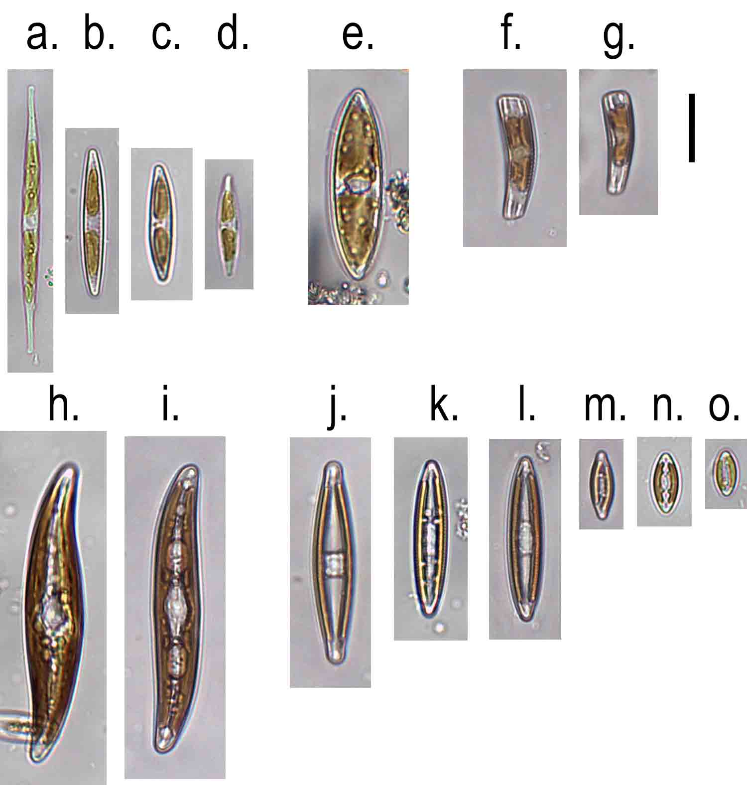
When I’m teaching people to identify diatoms, I start by telling them that there are two groups: the centric diatoms, characterised by at least one plane of radial symmetry, and the pennate diatoms, which have only longitudinal symmetry.  This is a good place for a beginner to start but reality is, inevitably, more complicated.  The two diatoms I describe in the previous post are, for example, both centric diatoms but it is difficult to discern this from the photograph, because their side views do not suggest any radial symmetry.  But, as I’ve said many times before, taxonomy and identification require very different approaches.

The basic centric/pennate dichotomy, however, is not just about outline, however convenient this is as a teaching aid for beginners.  Centric diatoms have a different mode of reproduction (oogamous) compared to pennates (isogamous) and also typically have many discoid plastids rather than a few plate-like ones.   I’ve illustrated one of the diatoms we saw at Deptford – Hydrosera triquetra – in the illustration at the top of the post, in order to delve deeper into this topic of shape as an identification aid.

The first thing we notice is that the valve face is not circular.  The literature describes it as “triangular” but, in reality, the shape is closer in shape to a star-of-David.  You could, however, take a pair of compasses and draw a circle that connected all of the valve angles, so you can see how this may be related to the more obviously circular diatoms.  The valve face is covered with a seemingly random array of coarse pores apart from the ends of three of the valve angles (termed “pseudocelli”) which appear to be plain, but which are, in reality, a mesh of very fine pores through which mucilage is secreted. This allows the cells to join together to form chains, or to attach to surfaces.  

Hydrosera triquetra is related to the other diatom I illustrated in the previous post – Biddulphia pulchella but whilst I can just about persuade you that Hydrosera is related to centric diatoms, it is harder to do this for Biddulphia, which is more obviously lanceolate in outline.  It also has psedocelli, which hints at a relationship with Hydrosera, and also has many small discoid chloroplasts (hinting at a relationship with the wider centric diatoms) but it sure ain’t round, by anyone’s definition.

The efforts of many taxonomists to shoehorn Biddulphia into the centric diatoms recalls Thomas Kuhn’s observations in The Structure of Scientific Revolutions – scientists approach data with strong preconceptions and outliers to the prevailing wisdom require ever more elaborate justifications to fit the pattern, something Kuhn described as “puzzle solving”.  Eventually, someone recognises that “normal science” is no longer viable, and proposes a new theory – a so-called “paradigm shift” (a phrase first used by Kuhn). 

In the case of Biddulphia and Hydrosera, the paradigm shift came in a 2004 paper by Linda Medlin and Irena Kaczmarscka who proposed a new class, Mediophyceae, for these “multipolar centrics” based on molecular sequence data, and that has now become the established classification (see “Who do you think you are?”).  Or, to use the language of Kuhn, we have re-entered a phase of “normal science”, at least until the next revolutionary biologist comes along with a better idea …

My picture was supposed to show Hydrosera triquetra growing on a vertical wall in Deptford Creek (with the bridge carrying the Dockland Light Railway over the river just visible in the background).  There are eight orders of magnitude difference between the Hydrosera cells (which are relatively chunky by the standards of diatoms) and the DLR bridge – which is a depth of field way beyond any camera could achieve.  Less impressive, from my point of view, is the appearance of a few mutant Hydrosera cells with five, rather than six, poles.  I finished the picture in rather a hurry as I wanted to post this before we headed off on holiday.  Sorry.

References

Ashworth, M. P., Nakov, T., & Theriot, E. C. (2013). Revisiting Ross and Sims (1971): toward a molecular phylogeny of the Biddulphiaceae and Eupodiscaceae (Bacillariophyceae). Journal of Phycology 49: 1207-1222.

Kuhn, T. (1962).  The Structure of Scientific Revolutions.  University of Chicago Press.

Medlin, L. K., & Kaczmarska, I. (2004). Evolution of the diatoms: V. Morphological and cytological support for the major clades and a taxonomic revision. Phycologia 43: 245-270.

Some other highlights from this week:

Wrote this whilst listening to:  Marika Hackman, following her gig at the Cluny in Newcastle.

Currently reading:  The Story of Art Without Men by Katy Hessel.  My daughter thought I needed educating.

Cultural highlight:  Marika Hackman in a packed Cluny.  Can’t help wondering why she’s not better known.  

Culinary highlight:  immediately prior to the gig, we ate at the Ship Inn, just across the road, which has all the attributes of a traditional Newcastle pub with football on big screens and “pub grub” on the menu, only the whole menu is vegan.  We ate their take on fish and chips – tofu wrapped in seaweed and then battered and deep fried – which rather proved my point that fish is algae-flavoured protein.

Dispatches from the city of migrants …

The journey to the location for this post started at the foot of the first “s” in the East Enders title sequence and ended beside the little blue squiggle at the bottom of the television screen, directly under the “a”.  I got off the Elizabeth Line at Stratford with no other plan other than that I had some time on my hands and headed roughly south, triangulating on glimpses of the O2 Arena and Canary Wharf as best I could.  I passed the splendid Italian Gothic edifice at Abbey Mills that houses the pumping station built as part of Bazelgette’s great sewer, walked alongside the muddy lower reaches of the River Lea, skirted numerous industrial estates and finally arrived, some two hours and nine kilometres later, at Island Gardens, a small area of green amidst the docklands sprawl, with a splendid view across the Thames to Wren’s Royal Navel College.  After crossing the river via the Greenwich foot tunnel, I emerged beside the Cutty Sark from where a short walk brought me to the blue squiggle that was my destination. 

That blue squiggle is Deptford Creek, scene of some posts at the end of last year (see “Floundering around in Deptford Creek” and “Throwing shapes …”) and I had come back to see how the project was progressing as well as finding some time to poke and scrape at more patches whose golden-yellow hue promised interesting diatoms.  The mudflats themselves looked much greener than I remembered, but it is hard to know if this is a genuine biological phenomenon or is just the random chance of two observations being different.  Today, however, I was more interested in what was growing around on the training walls at the edge of the creek rather than on the mud itself.  

My eye was caught straight away by bright green growths on the shelf on the wall beside the slipway.  Their distinctly felty texture made it easy to recognise the genus as Vaucheria although naming the species is a more difficult job.  We’ve met Vaucheria several times in freshwater (see “the pros and cons of cell walls …”) but it is also commonly found in brackish habitats.  My interest today was not in what species was growing, but in the way that it was helping to build a habitat within which a clump of moss and the shoot of a flowering plant were able to grow.  We saw this with happening with Rhizoclonium riparian during my previous visit (see “If only we looked …”); this time it is an alga from a different phylum, but we are looking at essentially the same phenomenon.

A patch of Vaucheria growing on the wooden training wall beside Deptford Creek, March 2024.  Note the moss (back right) and the shoot that have established themselves in the algal patch.  The patch is about 30 cm across.   The photo at the top of the post shows the training wall beside the slipway at Deptford Creek. 

Underneath this shelf, there was a distinct yellow-brown patch on the wall whose dominant constituent was Hydrosera triquetra.  I commented on its absence in a previous post (see “Deptford’s Heart of Darkness …”) but, in truth, the mudflat is not where I would most expect to find it.  It is a chain-forming diatom that seems to be associated with vertical surfaces and which can tolerate exposure. When individual cells are seen end-on (“valve view”), they are very distinctive, resembling a Star of David, with two overlapping triangles; however, the cells attach at these faces and the characteristic shape is not immediately apparent when seen in fresh material.  You need to focus carefully to understand the third dimension (see “Seeing with my fingers …”).  

Hydrosera triequetra in Deptford Creek, March 2024.  The left-hand image shows a patch growing on a wooden support (the central one visible in the image at the top of the post). The right-hand image shows a magnified view. The scale bar is 20 micrometres (= 1/50th of a millimetre) long.  

Hydrosera triquetra was first observed in the Thames in the early 1970s, and is widely-assumed to have been accidentally introduced, perhaps by a ship discharging ballast water.  Now it is widely-established.  I’m often sceptical of claims about “alien” species  but H. triquetra is sufficiently large and distinctive that it seems unlikely that it would not have been seen by earlier generations of microscopists at least occasionally.  Whether it justifies the term “invasive” is a moot point (see “Pens are too light …”).   That would require some evidence of wholesale changes in both the composition of the microscopic flora, and the way it functioned.  Personally, I love the diversity of modern London and am prepared to embrace a migrant diatom or two without feeling the need to apply pejorative adjectives.  

On the opposite side of the slipway, I noticed a seepage that, again, had a golden-brown hue suggestive of diatoms.  However, I could not see any Hydrosera growing amidst this assemblage, instead, there was a mix of Melosira varians and Biddulphia pulchella, a relative of Hydrosera, which lives in brackish water and forms zigzag colonies.  Motile diatoms such as Nitzschia sigma glided in and around these filaments.  The presence of Melosira varians is interesting.  I am fairly sure, based on examination of living cells, that these filaments are not one of the related species of Melosira that are more tolerant of salinity. That a freshwater species with only mild affinity for salt is thriving here supports some observations of higher plants on the slipway made by Nick and Andy from Creekside Discovery Centre. They noticed that these, too, are mostly not especially salt-tolerant plants, leading them to think that the upper slipway represents a freshwater tidal marsh, rather than a salt marsh, and that it is the result of fresh water from the Ravensbourne being lifted above the denser salt water that pushes into Deptford Creek on each tidal cycle.  A freshwater species tolerant of some salt rubbing along beside a brackish species that can cope with an occasional dose of freshwater seems like a good metaphor for this part of London where people from every part of the world live side-by-side.

Another seepage beside the slipway at Deptford Creek, this time with a mix of Melosira varians (right) and Biddulphia pulchella (left). The scale bar is 20 micrometres (= 1/50th of a millimetre) long.

Some other highlights from this week:

Wrote this whilst listening to:  vintage Jefferson Airplane and, for contrast, Kendrick Lamar’s To Pimp A Butterfly.

Currently reading:  The Secret Diaries of Charles Ignatius Sancho by Patterson Joseph, a novel that is, by coincidence, about 18th century migrants in London.

Cultural highlight:  Lines, a play at the Crucible in Sheffield exploring injustice in Palestine and Uganda.  Rather challenging.

Culinary highlight:  Seven course tasting menu at Prashad, an Indian vegetarian restaurant  in Drighlington, just south of Leeds

Call me by my name …

One of my very earliest posts addressed a very basic problem in the study of microscopic organisms: knowing the identify of whatever you are looking at (see “All exact science is dominated by the idea of approximation …”).  The germ for this post came from a quotation from Bertrand Russell that said that, when you counted anything, you had a good idea of what constituted whatever it was you were counting.   If you were counting oranges, you knew to exclude apples, obviously, but also tangerines and satsumas, which may look similar but are genetically-distinct.  That’s not always so straightforward for microscopic algae, as I’ve discussed in many posts, but it is a core, largely unspoken, assumption.   I’ve also emphasised the distinction between the sciences of taxonomy and systematics, and the craft of identification (see “Disagreeable distinctions …” and, more recently, “How to identify diatoms from their girdle views …”).  As an ecologist, I need to use the outputs from taxonomy but, at the same time, I am constrained by time, equipment and, indeed, by the knowledge that I can readily access, in my ability to do this.  

I’ve tried to encapsulate this as two overlapping triangles in the diagram below.  The reason why we cannot name a specimen distils into one of two reasons: limitations of knowledge (represented by the blue triangle) or practical limitations such as the quality of the microscope or the way that the specimen presents on the microscope slide.   So every identification challenge sits somewhere on the gradient from left to right: we are either limited by available knowledge or practicalities.

The challenges involved in identifying diatoms (and other microscopic organisms) fall into two types: the quality of the knowledge that we have available, and our ability to link this knowledge to the specimen we see under our microscope.  

Knowledge limitations encapsulates everything that is known at the time that the specimen is being analysed, and will include the following: 

  • The species within a genus may have not yet been fully discriminated and described for the region you are studying;  
  •  There are limitations associated with light microscopy which means that the full range of genetic variation within a complex of species cannot be resolved (“crypticity”).
  • The literature for that genus is dispersed in numerous journal articles rather than gathered in a single accessible volume.  In theory, we can access these via Google Scholar or Web of Science, but some will be behind paywalls.  Sometimes, knowing where to look is, itself, the problem.
  • The keys in taxonomic works are often poor (which inhibits identification by logical reasoning) and/or there may be insufficient images (inhibiting pattern recognition) and there is often insufficient focus on critical differentiating characteristics.  

Next, there are problems that relate to the practicalities of identification, irrespective of the knowledge that is available:

  • Individual cells do not present in an optimal manner (e.g. as girdle views in the case of diatoms) or key diagnostic criteria are absent (reproductive organs are necessary to identify species of filamentous algae such as Spirogyra and Oedogonium) or the taxon is sufficiently sparse in the sample that the full range of morphological and diagnostic characteristics cannot be discerned.
  • Equipment available is not sufficiently sensitive.   A common problem when instructing students is that microscopes used in university teaching laboratories have a lower specification than those used for research, so the students often cannot see the same level of detail that the textbooks assume.  A twist on this general theme is that the camera on a microscope used for routine analyses cannot capture the level of detail required to allow someone else to identify a specimen when you email them an image. 
  • The observer is unaware of recent literature or is working under time constraints that prevent a full literature search.   This is similar to the third point in the “knowledge” section, reflecting a second facet: problems arise not only because specialists are no longer prepared to write detailed monographs that gather together the literature on a genus in a region, but also because time and budget limitations mean that the alternative – thorough interrogation of a very diffuse literature  – is not always possible. 
  • Faulty memory links a particular set of morphological properties to the wrong name.  Mistakes, in other words.  There is a danger that our recollection of the properties associated with a particular name drifts over time, and needs regular calibration.

In reality, I often have to make a judgement about the value of any extra information that I can glean from a sample, relative to the effort I will need to in order to unlock this information.   There is a basic scientific curiosity in me that wants to know the identity of an alga I’ve never seen before, and to understand the diversity within a habitat better, but I’m also often working under pressure and spending time trying to name a single cell whose identity eludes me will add very little to the interpretation based on the other 299 cells I’ve already examined and named.  On the other hand, if there was a single volume work on the Fragilaria of Europe (for example), the marginal effort required to make that extra determination might be reduced so that I can pluck the book from my shelf, flick through the pages, name it and move on.  The danger of this lies in the first point I made – about incompleteness in our basic understanding of the variety within most genera – and means that a monograph may, itself, not be sufficiently comprehensive.  “Hard cases” will be force fitted to available descriptions, perpetuating the longer term problem of fitting morphology-based descriptions to the biological species concept. 

There was a time, not so long ago, when there was a general belief that there was a finite number of cosmopolitan species of freshwater diatoms, and an unspoken assumption that all could be differentiated with light microscopy.  Over the last thirty years, those ideas have been superseded as forms once regarded as a single species have been fragmented into a multitude of distinct entities.   No-one seriously doubts that there are many more species than diatomists a generation older than us had contemplated, but one side-effect is that the capacity of Linnaean taxonomy to serve as helpful “tags” that ensure consistency in identification between scientists is close to breaking-point.   The craft of identification depends to a large extent on the willingness of taxonomists to invest time not just in expanding our knowledge but also in gathering this together into practical resources.  That’s now become such a Sisyphean task that few seem to want to take it on.  Yet, not having such resource creates problems of its own, not least in contributing to a sense amongst bureaucrats that diatomists are better at talking the talk than walking the walk, in terms of their ability to contribute to answering the pressing environmental challenges of our day. 

Some other highlights from this week:

Wrote this whilst listening to:  New Energy by Four Tet.

Currently reading:  Still on Amy-Jane Beer’s Flow.   Her explorations of British rivers includes a snorkalling trip in West Beck / River Hull which I wrote about in “Past Glories …”.

Cultural highlight:  A World in Common: Contemporary African Photography – an exhibition at the Tate Modern in London. 

Culinary highlight:  When I first started this post, I was in Belgrade, eating traditional Serbian food in the Tri Seseria restaurant in Belgrade’s old town.  That was in an unseasonably warm October, and the unfinished post has sat on my laptop for almost three months awaiting completion.   

If only we looked …

We stay in Deptford Creek for this next post, moving a short distance upstream from the mud where I encountered diatoms and euglenoids and looking, instead, at the walls that define the creek’s channel.  What should I call these walls?  Not exactly “embankments”, certainly not “levees”, nor “quays”, maybe “revetments”.  I came across some old papers, written by civil engineers, which referred to these as “training walls”, suggesting that the errant creeks need some firm discipline to keep them from wandering too far into the human realm.  I like to think that we live in more enlightened times now, that rivers are better left to define their own channels.  However, I suspect the options for “rewilding” a tidal stream in central London are rather more limited than in the parts of the country where I usually work.  I’ll just say “walls” from now on.

You can see from the photos that there is a distinct green growth on this wall, starting about 90 cm above the mud.   A closer look at this revealed it to be a mat of fine green interwoven filaments, each about 25 micrometres (1/40th of a millimetre) in diameter.  This is Rhizoclonium riparium.  Superficially, it resembles Cladophora glomerata, the common blanket weed of freshwaters, but the filaments are narrower and unbranched, apart from the occasional rhizoid (see lowermost image in the plate below).  This is a very common species on hard surfaces in brackish intertidal habitats.  Here, in Deptford Creek, it is doing more than just adorning the walls.  It is also helping to build the habitat.

Rhizoclonium riparium from the walls of Deptford Creek, November 2023.  Scale bar: 20 micrometres (= 1/50th of a millimetre).  The picture at the top of the post shows the walls from where the alga was collected.

We’ve seen examples of algae as “ecosystem engineers” in the past (see “Landscape architects …” and “How to make an ecosystem”).  This example is different in that it involves a green alga (rather than a cyanobacterium) and it is on a vertical, rather than a horizontal, surface.  Each day, the tide rises and falls twice, submerging the algae in salty water.   The interwoven mat of filaments also enables it to trap any small particles suspended in the water so gradually, over time, this mat of algae takes on some of the characteristics of soil.   Any seeds that arrive, either in the water or from the air, may also get trapped in this mat and, so long as they are tolerant to saline conditions, will germinate.  The final picture in this post shows some of the plants growing in this algal “soil”.  I think I can see pellitory-of-the-wall (Parietaria judaica) in there, but there are other species too.  These will not be submerged during every tidal cycle, but the algae mats and their associated “soil” will act as “wicks”, carrying water up to the plant roots higher on the wall.

David Attenborough, another London resident, once said: “Looking down on this great metropolis, the ingenuity with which we continue to reshape the surface of our planet is very striking. It’s also very sobering, and reminds me of just how easy it is for us to lose our connection with the natural world”.  The view from Deptford is one of algae also reshaping the surface of a city and of connections with the natural world right on our doorsteps, if only we looked …

Plants growing in the Rhizoclonium mats on the walls of Deptford Creek.

Some other highlights from this week:

Wrote this whilst listening to:  my alternative Christmas playlist, reacquainting myself with old favourites such as Slow Club’s It’s Christmas and You’re Boring Me, Half Man, Half Biscuit’s It’s Cliched to be Cynical at Christmas and Phoebe Bridger’s If We Make it Through December.

Currently reading: still on Jerusalem: the Biography, by Simon Sebag Montafiore.  A well-written book but unrelenting in the descriptions of bloodletting over the years.  Essential reading for anyone who wants to understand the present conflict.

Cultural highlight: Julia Brook’s exhibition What Is It That Will Last at Abbot Hall in Kendal.  An artist with a “deep and immersive relationship with the landscape” … literally, because she, too, has to study tide tables closely before venturing forth.

Culinary highlight: Tasting menu at The Old Stamp House in Ambleside.   Michelin-starred restaurant whose brilliant food is sustainably sourced from the locality.  One of the best meals I’ve eaten this year.

Floundering around in Deptford Creek …

“Fieldwork” usually involves pointing the car towards the countryside and leaving all the travails of urban living behind for a day or two.  I like to stand amidst glorious scenery sucking pure air into my lungs and reflecting on how lucky I am to do the work I do.  But all that changed earlier this week.   My destination was not the Pennines or the Lake District but a muddy creek in Deptford surrounded by blocks of flats, old industrial premises and a viaduct that carried the Docklands Light Railway into the badlands of south London.  

Just out of sight on  the right of the picture at the top of the post is the low wooden building of the Creekside Discovery Centre, a charity that promotes urban wildlife.  My involvement here is part of a citizen science project that will help the staff at Creekside raise awareness of the microscopic algae that inhabit Deptford Creek.  It is co-ordinated by Susi Arnott, a filmmaker, whose film Moon, Mud and Microbes I took to Green Man in 2021 (see “Cover versions …”).   I

The visit had to be closely-calibrated to coincide with low tide, so that we had as much time as possible wallowing on the mud looking for algae.  I’m somewhat out of my comfort zone, but we soon spotted a rich chocolatey-brown patch on the mud surface that looked extremely promising.  This would have been submerged at high tide, and had probably been exposed for five or six hours by the time we visited.   I scraped off some of the growth to look at later, although we also tried a variant of the “lens tissue” method for sampling (see “Eyes wide open in Cassop’s muddy fringe …”).   My sample turned out to be full of a large sigmoid Nitzschia, cells of which glided to and fro across the field of view as I peered down my microscope.   I’m going to be circumspect about naming the species because I’m not familiar with brackish water diatoms and there are a number of possible candidates, but the genus is unmistakable.  

The patch of diatoms growing on a low shelf on the embankment at the right hand side of the image at the top of the post.

I felt more at home standing in the middle of the creek itself.   The bed here is rocky rather than muddy and the water, at low tide at least, is fresh.   There are some distinct diatom patches on some of the stones here too, and I picked some up using a toothbrush to look at later.  Again, I’m tempted to name them but, with the exception of Rhoicosphenia abbreviata (which is both distinctive and can tolerate brackish conditions), I’m reluctant to do so without having looked at cleaned frustules.   There are species of Navicula, in particular, which have similar outlines and dimensions to common freshwater species but which, on close inspection are something else altogether (see: “More diatoms that like cold weather …”).

The sigmoid Nitzschia growing on mud surfaces in Deptford Creek, November 2023.   Scale bar: 20 micrometres (= 1/50th of a millimetre).  

More diatoms from Deptford Creek.  a. – d. Nitzschia spp.; e. Tryblionella sp.; f.g. Rhoicosphenia abbreviata; h., i. Gyrosigma; j. – o. Navicula spp.   Scale bar: 20 micrometres (= 1/50th of a millimetre).  

There was another diatom with a sigmoid outline in this second sample.  I wish I knew what advantages this curved outline confers on an organism because this is the not the first time that I have seen two sigmoid genera, quite distantly related, living so close together.  This particular organism is Gyrosigma.  Whilst I need cleaned material to be sure of what species of Gyrosigma I’ve got, it is easier, ironically, to differentiate the genus from Pleurosigma when looking at live material because the plastids (chloroplasts) have different structures (see “Microscopic monsters in mud …”).   Pleurosigma has two or four ribbon like plastids which have a convoluted form whereas the two plastids of Gyrosigma hug the valve outline.  Despite outward appearances, these two genera belong to different families.

Both samples were dominated by diatoms that can move around, reflecting the shifting, unstable nature of the habitat within which they live.   When viewed under a microscope we see this as horizontal movement but, out on the mud of Deptford Creek, this will be in three-dimensions, including a daily “commute” from the deeper parts of the mud to the surface in phase with the tides.   We see them gliding serenely across a glass slide but the process is more like a climber ascending a tight “chimney” on a rock face – first one, then another of the diatom’s two raphes will be engaged, and sometimes both together.  Talking of commuting, the sun was starting to descend on Deptford Creek and I had a train to catch.   I emerged from the tiny oasis that the Creekside Discovery Centre nutures and joined the stream of people heading towards Deptford Station to start the journey back home.

If you are wondering why the word “floundering” appears in the title, look at the picture below.  We were walking up the creek when we noticed this fellow who seemed to have got stuck in the shallow water of the creek.   He didn’t seem too alarmed by our presence (or maybe he just didn’t have too many options for escape – flounder body language is not something I know much about), allowing me to get my camera into the water to take some pictures.   

Some other highlights from this week:

Wrote this whilst listening to:  Patti Smith’s Horses and west coast cool jazz on Chet Baker and Art Pepper’s 1956 album Playboys

Currently reading: Colm Tóbín’s Homage to Barcelona

Cultural highlight: Installation by Céline Condorelli, Artist-in-Residence at the National Gallery in London, featuring a rug whose abstract patterns were based on the patterns of pigments on pictures from the gallery’s collection viewed under a microscope.  The title Pentimenti (the corections) comes from the use of imaging technologies to discern the changes artists make during the process of composition. 

Culinary highlight: nut roast and trimmings: Sunday lunch cooked by our daughter, followed by a rather good vegan sticky toffee pudding.

Part of Pentimenti (the Corrections) by Céline Contorelli in the National Gallery, November 2023.  

Type dilemmas …

The diatom genus Achnanthidium frequently appears in this blog.  It is widespread in freshwaters, particularly in cleaner water, and telling the many species apart is one of the banes of my life.   To cut a long story as short as is possible, members of this genus are typically between 10 and 20 micrometres (1/100th and 1/50th of a millimetre) and push the capabilities of light microscopy to the limit.  Until recently, it was usually treated as part of a larger genus Achnanthes, and most specimens were treated as “Achnanthes minutissima”, a species first described in 1833 by the German botanist Friederich Kützing.  Even by 1930, we find only three species included in Hustedt’s  Die Süsswasser-flora Mitteleuropa.  The widespread availability of scanning electron microscopes revealed a lot more variation within Achnanthes minutissima (or Achnanthidium minutissimum, as it has been known for the last 30 years or so), leading to the description of many new species.  As many of their properties are difficult to resolve with a regular light microscope, identification is always challenging.  

As these taxonomic studies inevitably mean that the historic “broad” Achnanthidium minutissimum is “split” to create new species, the convention when describing a new species is to refer back to the original description and the “type specimen” and explain how the new species differs from this.  This means that several papers have been published recently which examine Kützing’s original publication.  I’m usually focussed on the finer points of diatom morphology that these papers discuss, but last week something odd struck me about Kützing’s description of “Achnanthes minutissima”.  The specimens he describes were growing “on Zygnema with Exilaria crystallina in a ditch near Aschersleben, in June.”   Aschersleben is a town in Saxony in central Germany about 80 km from Nordhausen where Kützing lived for much of his life but, this detail apart, nothing else in this account of the habitat rings true.   First, the drawing shows the diatom growing epiphytically on an alga that lacks the two star-shaped chloroplasts that characterise Zygnema (see photos in “A day out in Wasdale …”).  Second, Zygnema and relatives typically produce copious mucilage and, as a result, have few epiphytes (the exception would be a dead or dying filament).  Third: Exilaria crystallina is an old name for a diatom whose name has changed several times but which was called Ardissonea crystallina until very recently when it was transferred to Synedrosphenia.  It is, however, a brackish and marine species, not one that is found in freshwaters.  It is also, by diatom standards, an enormous species (up to 0.7 mm, according to the literature).  It would also be very surprising to see this species growing alongside Zygnema and Achnanthidium, both of which are intolerant of brackish conditions.

This is not intended as a scathing critique.   What appears in the illustration is certainly an Achnanthidium species – a small cell with a bent girdle attached to a surface by a short stalk.  It is everything else in the description that set me scratching my head.  Kützing was working 200 years ago with much less sophisticated equipment and, perhaps more importantly, without the network of supportive peers that modern biologists take for granted.  I’m more surprised that, amidst all the attention this description has received in recent years, no-one seems to have commented on the incongruity of the habitat details.   There is, it seems, an elephantine diatom (Syndrosphenia crystallina) in the corner of the room and no-one has noticed.

Reference

Lobban, C. S., Ashworth, M. P., Camacho, T., Lam, D. W., & Theriot, E. C. (2022).  Revision of Ardissoneaceae (Bacillariophyta, Mediophyceae) from Micronesian populations, with descriptions of two new genera, Ardissoneopsis and Grunowago, and new species in Ardissonea, Synedrosphenia and ClimacospheniaPhytoKeys 208: 103.

Some other highlights from this week:

Wrote this whilst listening to:  Grace Petrie, folk/protest singer

Currently reading: The Slow Road to Tehran, an account of a journey from London to Tehran via Sudan by Rebecca Lowe.   

Cultural highlight:  the 1983 film Yentl, starring and directed by Barbara Streisland.  

Culinary highlight: home-made lemon meringue pie.  Comfort food

Dancing to different rhythms …

Another river, another algae-strewn boulder.   This time, we’re at the River Irt, also in Cumbria, and the green algae, under the microscope, are mixed growths dominated by Mougeotia, Zygnema and Spirogyra rather than the monoculture of Oedogonium that we saw in the River Cocker.  You can also see brown patches of Tolypothrix on the boulder in the foreground.

The surprise, for me at least, was the presence of conjugation tubes in the Mougeotia, bearing in mind my comments in the previous post.  Did I or did I not write that “filamentous algae don’t see the need for sexual reproduction” only a few days ago, and am I or am I not now staring down my microscope at a filamentous alga doing just that?  There is no obvious trigger for this: the generally-cited hypothesis is that the zygospores produced as a result of conjugation are resistant to adverse conditions; however, cool stream water in a northern English river is really a quite benign environment in which to grow.  I expect to find lush growths of Mougeotia and relatives here and in other Lakeland rivers throughout the winter.   

I’ve tried to capture some of this wanton behaviour in a picture that shows some of the stages that I could see.   In the top left you can see some “papilla” emerging from the cells of adjacent cells then, in the centre, two papillae have met and fused to form a conjugation tube.  What will happen next is that the cytoplasm of the two cells joined by the tube will migrate into the tube and fuse to form the zygote; however, I could not see this in my material.  It is thought that there is some cell-to-cell signalling so that papillae grow from adjacent filaments and meet in the middle, but the nature of this signalling seems still to be a mystery. 

Mougeotia from the River Irt, October 2023.  A conjugation tube has formed between the two filaments at the centre of the image, and tubes have also started to grow from the filaments in the top left corner.  The filaments are about 25 micrometres (1/40th of a millimetre) in diameter.  The photograph at the top of the image shows submerged boulders smothered with Mougeotia and Tolypothrix on the bed of the River Irt.  

In the absence of zygospores in the Mougeotia from the River Irt, I have included two images produced by Chris Carter.  In both of these you can see the zygospore forming in the middle of the conjugation tube.  This is in contrast to what happens to Spirogyra, a close relative of Mougeotia, where the cytoplasm from one filament (designated “male”) moves right through the conjugation tube and fuses with the “female” in her own cell.

The zygospore has a very tough cell wall and, as a result, is very resistant to adverse conditions.   Having a form of sexual reproduction that is triggered by the environment seems strange but that’s partly a matter of perspective.  The angiosperm lifecycle – pollen, flowers, seeds and all that caboodle – is an adaptation to a perennially unfavourable environment, when seen from an alga’s viewpoint, so the question of when it is most useful to have sex shifts from merely coping with occasional adversity to a broader need to be in phase with seasonal cycles.  We are also terrestrial organisms, long conditioned to watching for seasonal prompts in the nature around us, so it is difficult for us to understand the different rhythms to which riverine algae dance.     

A zygospore of Mougeotia scalaris from Yardley Chase, Northamptonshire, photographed at three focal planes by Chris Carter.

Zygospores of Mougeotia parvula from Malham Tarn, photographed by Chris Carter.

Reference

Permann, C., Herburger, K., Niedermeier, M., Felhofer, M., Gierlinger, N., & Holzinger, A. (2021). Cell wall characteristics during sexual reproduction of Mougeotia sp.(Zygnematophyceae) revealed by electron microscopy, glycan microarrays and RAMAN spectroscopy. Protoplasma 258: 1261-1275.

Some other highlights from this week:

Wrote this whilst listening to:  The Ghost of Tom Joad by Bruce Springsteen, celebrating our purchase of tickets to see him next May.

Currently reading: Primary Colours, novel by “Anonymous” (Joe Klein) based on Bill Clinton’s 1994 campaign.

Cultural highlight:  A visit to the new Faith Museum at Bishop Auckland

Culinary highlight:  meal from Sattvic Ahara, a new Indian vegetarian takeaway in the next village.

The view across Derwent Water from Keswick on the day before my October fieldwork.

The long wait is over …

I have not been to my regular haunts in the western Lake District for two months now.   We usual collect samples at two month intervals but our August trip was compromised by the wet weather we had during the latter part of the summer.  You can see the evidence for this in the graph below, showing how river levels fluctuated.   I should point out that, for personal safety reasons, we can only go out when the river is low so even the values during the latter part of August would have made wading problematic.  Samplers, it was once pointed out to me, are part of the benthos, not part of the plankton.  Also, values on the river level graph are recorded at the gauging station, and will not be the same at the point where we sample.  We have learned, over the years, to make a mental adjustment to get a sense of what conditions will be like when we arrive.

By early September, however, levels were low once again but, annoyingly, I had other commitments, including the course I wrote about in “Cyanobacteria inside their comfort zones …”.   Anticipating river conditions based on weather forecasts and juggling with other diary commitments is all part of the job.  This particular trip has been harder to organise than most.   I have had to make and cancel hotel reservations on three separate occasions, and on a fourth, I drove across and stayed overnight, only to find the rivers were higher than I had hoped when I checked the hydrograph first thing in the morning.  

River levels in the River Cocker (Scale Hill) from 1 August to the time of writing.   Data from www.riverlevels.uk.   Values are averages for each day and the dashed line is the level on the day of my visit.  The photograph at the top of the post shows Oedogonium growing on submerged stones in the River Cocker at Low Lorton.

When I finally got to the River Cocker, I was greeted by the sight of lush growths of green algae across the riverbed.  This turned out to be a near-monoculture of Oedogonium, which we last encountered in the River Derwent earlier in the summer (see “Borrowdale landscapes …”).  It is a very common genus, albeit one that is very hard to identify to species and also one for which generalisations about ecology are difficult.  

What I can say, with some confidence, is that the quantity of algae present in these streams waxes and wanes in a predictable manner, with highest values recorded in winter, but it is not so easy to say which species will proliferate on any particular occasion.  That also gives us a clue about the possible reason for the patterns that we see: because the biomass fluxes happens at a number of sites across several catchments, irrespective of which species are present, it must be driven by external factors that are common to all of these sites.   And because the fluxes are most extreme in rivers that are downstream of lakes (as is the case for the Cocker), we suspect that temperature plays a role.  Because water has a high specific heat capacity, Crummock Water acts as a huge “heat pump”, making the water in the Cocker ever so slightly warmer during autumn and early winter than is the case in nearby rivers that do not drain out of lakes. 

Filamentous algae are well placed to take advantage of this growth as they are simple and straightforward photosynthesis machines.  Sunlight is trapped by their chloroplasts and converted into the building blocks of cells which divide mitotically, allowing biomass to accrue without the complications faced by more sophisticated organisms.  There is a certain amount of phenological control in some filamentous algae, but this is mostly concerned with the onset of sexual reproduction (see “The intricate ecology of green slime …”).  Most of the time, filamentous algae don’t see the need for sexual reproduction (see “Tales from the splash zone …”) so the Oedogonium in the River Cocker is free to take advantage of the slightly warmer water compared to nearby streams, and convert as much of the late autumn sunlight as possible.  Meanwhile, the bugs that normally graze away any algae that cover submerged stones are dancing to the tunes played by their own internal clocks and lack the capacity to increase as quickly as their food supply.  The result is the green riverbed that I saw in the River Cocker when I visited. 

Oedogonium filaments from the River Cocker, October 2023.   The lower image shows a cell with a number of cap cells.  Scale bar: 20 micrometres (= 1/50th of a millimetre).

My comment about Oedogonium being able to accrue biomass without the complications faced by larger organisms needs a little qualification, because not every cell is able to divide.  The photograph above shows a cell with a fine collection of “caps”, demonstrating that it has divided numerous times.  There is, in other words, a first tentative step towards specialisation of cell function and, from this, we may also infer some redistribution of photosynthesis products along the filaments.  

Some of these “cap cells” were conspicuously brown compared to the cells on either side.  This is quite a common sight but I have found nothing in the literature that may explain what is going on.  The colour is suggestive of ochre and my working hypothesis is that these cap cells are particularly metabolically active, requiring the chloroplasts in the cell to work harder than the cells on either side. This will mean that these cells evolve more oxygen and this, in turn, will mean that iron and manganese in the water are more likely to precipitate out.  The lower photograph shows one of these brown cap cells colonised by diatoms (Gomphonema) and filamentous bacteria.   Oedogonium often has a substantial payload of epiphytes but not usually concentrated in a few locations.  Once again, what is it about the cap cells in particular that makes this a good location for other algae?   The literature is silent. 

More Oedogonium filaments from the River Cocker, October 2023, this time showing iron/manganese preciptitation around cap cells and associated epiphytes.  Scale bar: 20 micrometres (= 1/50th of a millimetre).

I always leave my regular sampling locations curious about what I will see next time I visit.  This time, however, this curiosity is leavened with a sense of trepidation as the long-term weather forecasts seem to suggest we are in for a wet autumn (driven by the El Niño in the western Pacific).  As a result, I am also anticipating spending more time over the next few weeks poring over the hydrographs and weather forecasts trying to predict when river levels will be low enough to pull on my waders and get back into the river.  Uncertainty will be the only certainty in my life over the next few weeks …

Some other highlights from this week:

Wrote this whilst listening to:  Frankie Archer, who blends traditional folk music with electronica.  I saw her on Later … with Jools Holland a couple of weeks ago and then found that she was playing at Darlington Library a few days later.   Her haunting melodies have stayed with me …

Currently reading: Qian Zhonghsu’s Fortress Beseiged.  Classic Chinese novel set on the eve of the Sino-Japanese war.

Cultural highlight:  A Ken Loach double-bill: first, a stage adaptation of his film I, Daniel Blake at the Gala Durham, then his latest film The Old Oak at the Tyneside Cinema.  The latter uses several locations in Co. Durham including Blackhall Rocks (see “County Durham’s tropical seashore”)

Culinary highlight:  vegetarian tasting menu at Rebel in Heaton.

Cyanobacteria inside their comfort zones …

Last week saw the second presentation of the FBA’s algae identification course for 2023, with another dozen keen participants being introduced to the intriguing world of freshwater algae.  The week starts with sampling in the vicinity of the FBA’s laboratory, first at Windermere itself (see “A hitchhiker’s guide to phytoplankton …”) and then, the following morning, at other sites in south Cumbria.  I usually join the group heading to Cunsey Beck, which flows from Esthwaite Water to Windermere but heavy overnight rain meant that river levels were too high for safe wading.   Instead, we joined Allan Pentecost on a trip to White Scar Quarry, right at the south-eastern tip of the Lake District.   It is an area that Allan knows very well and I always enjoy visiting with him because I learn a lot (see, for example, “Love and sex in a tufa-forming stream …”).

Just at the edge of the quarry, a small seepage flows across a wide bedding plane and, Allan pointed out to us, an impressive range of Cyanobacteria, green algae and moss co-existed side-by-side.   If you look at the photograph at the top of the post, you can see the seepage emptying onto the bedding plane just above the “d.”.   At low flow conditions, there is just a gentle trickle of water running in a narrow band approximately at the centre of the picture.   However, when there is more water coming down the quarry, then the water spreads out further and is also augmented by rainfall landing directly on the bedding plane.  The result is a zonation, roughly akin to what would be seen on a rocky shore, albeit not for seaweeds.  

Following this from the left we have:

  1. The driest area, dominated by Nostoc commune whose ecosystem-building capabilities in otherwise adverse environments I’ve described in earlier posts (see “Landscape architects …” for the most recent of these);
  2. An only occasionally wetted area dominated by dark brown mats of Scytonema.  The colonies described in “Poking around amongst sheep’s droppings …” came from very close to here and will give you some idea of what to expect;
  3. A slightly wetter area has reddish mats of filamentous cyanobacteria. Schizothrix, Phormidium and Homoeothrix all feature here.  An example of a Phormidium growing at the air-water interface (and, therefore, presumably tolerant of desiccation) is described in “Fieldwork notes, August 2021”)
  4. The central zone is almost permanently wet and here there is a distinct growth of the green alga;
    Mougeotia, a very common alga in this part of the world.  Recent posts which mention this genus include “Something, somewhere, just for a moment …” and “The man who stares at algae …” (both, incidentally, describe the interplay between green algae and Cyanobacteria in Lake District streams).

From here, the sequence is reversed except, at the right-hand side of the picture frame (e.) we did not see more Nostoc commune but, instead, patches of Rivularia haematites and bright patches of the moss Philonotis fontana.  I last wrote about Rivularia in “Building landscapes …”, based on another excursion that Allan led, this time in the Malham area of Yorkshire rather than in the Lake District.   I wonder, in retrospect, if the moss is, itself, a result of the ecosystem-building properties of Nostoc that I mentioned above and also wrote about in “How to make an ecosystem (2)”.   If I had teased apart those Philonotis clumps, would I have seen colonies of Nostoc lurking at the bottom, perhaps?   We hear a lot about all the problems Cyanobacteria cause in the Lake District at the moment so it is useful, once in a while, to remind ourselves that Cyanobacteria helped to build this wonderful landscape in the first place.

Microscopic views of the zonation at White Scar Quarry, Cumbria, September 2023.  a. Nostoc commune; b. Rivularia haematites; c. Phormidium sp.; d. Mougeotia sp.   No scale bars, I’m afraid, as the images were grabbed ad hoc while I was teaching, but other posts referenced should all have indications of size.

Some other highlights from this week:

Wrote this whilst listening to:  Africa Express Presents … The Orchestra of Syrian Musicians and Guests.  

Currently reading: Maggie O’Farrell’s The Wedding Portrait, set in Renaissance Italy.

Cultural highlight:  Exquisite Korean-American film Past Lives directed by Celine Song.  A love story mostly set against the Manhattan skyline.

Culinary highlight:  It may seem like a minor achievement, but I made the best shortbread of my life this week, following the Ayrshire Shortbread recipe in Cerys Matthew’s cookbook, Where the Wild Cooks Go ….  The vegan haggis that I made from her recipe also went down well a couple of weeks ago.

Patches of Philonotis fontana on the bedding plane at White Scar Quarry, September 2023.

CSI: algae …

My go-to analogy for the type of freshwater ecology that I do is of being a “general practitioner” (family doctor) for lakes and rivers.  I check my “patients” at regular intervals and offer them “prescriptions” to help them recover.  Just occasionally, however, situations demand a different analogy, that of the hard-boiled detective, roused from semi-slumber by the shrill ring of a telephone and summoned to the scene of a crime.  Sometimes there is even blood involved, or at least so it seems.  I am the one who announces to the protagonists assembled in the drawing room that it is not blood at all.   It is … algae.   

Freshwater ecologists have a fairly good idea of what algae grow where, how they wax and wane over the course of the year and how human activities can alter these patterns.  However, we also know that there is a limit to the extent that we can predict, and that there are many occasions when a single species can proliferate to the extent that members of the public can see, and be concerned by, the changes in a local water body.   In most cases, these growths are benign but there are situations – cyanobacteria blooms being the best known – where there are serious implications for human and animal health.

The algae that can cause these problems can come from any one of several groups such that the initial diagnosis necessitates a good general background in algal taxonomy.  We try to provide this background on the FBA course on freshwater algal identification as many of the participants work for regulators and water companies and are “first responders” to calls from concerned members of the public.  On the final day, Bill Brierley gives a talk on “incidents” based on experiences from his long career in the Environment Agency.  

What is striking from this talk is that representatives of all the main groups of algae [see Origin story … ] can be the culprits, along with a few groups that are not algae but which can be confused with algae.   Quite a few have been covered in this blog over the years.  Here’s a quick summary, along with some links.

In the Prokaryotic domain, we have the Cyanobacteria as the serial villains of the freshwater world (see: “A hitchiker’s guide to phytoplankton …”) but other groups can also cause problems.  “There will be blood …” describes a bloom of Chromatium, a purple photosynthetic bacterium, which turned a pond close to where I live an alarming red colour.

However, red-coloured blooms can come from a number of Eukaryotic algal groups as well.  Euglena sanguinea from the Euglenophyceae (Protozoa), in particular, can have a similar effect in standing waters and slow-moving rivers.  One of the key lessons that emerged from Bill’s talk was the need for samples to be examined by a biologist with a broad understanding of algae because it would be easy to jump to the wrong conclusion if a bloom was only viewed from the shore.

A bloom of Euglena sanguinea in a pond in eastern England.  Photograph: Bill Brierley.  The photograph at the top of the post shows an unexplained proliferation of green algae in a river in Cumbria.

Several problem-causing organisms can be found in the Chromista (see “Unlikely bedfellows …“).  The Haptophyta includes some toxin-producing groups including Prymnesium and Chrysochromulina both of which are more common in brackish and marine waters but which can extend inland, especially when salt concentrations are artificially elevated.   In the summer of 2022 a bloom of Prymnesium parvum had catastrophic effects on the River Oder in Poland and Germany, killing about 360 tonnes of fish.  In this case, discharges from salt mines in Poland were responsible for raising the salinity of the river and this, combined with high concentrations of nutrients and low flows, created a lethal cocktail. 

Left hand images: Prymnesium parvulum (note the short, rigid “haptonema, cells about 10 micrometres long); right hand image: Chrysochromulina braunii.  Photographs: Bill Brierley

Both of these genera are widespread in low-lying and brackish lakes in Norfolk and Lincolnshire and the expectation is that the favourable conditions under which they thrive will become more common as climate change continues, leading to more problems in the future.  

The other group of Chromista that are often associated with problems are the diatoms.  Although some marine genera do produce toxins, there is scant evidence for fish kills that are directly attributable to diatoms in freshwaters.  It is more common for mass growths on the beds of rivers to attract attention from passers-by.  The diatom Didymosphenia geminata is a repeat offender in this respect.   Although regarded as an invasive species in some parts of the world, it is native to the UK with records extending back over 150 years (see “Journey to the headwater of the River Coquet”).  Nonetheless, when conditions are right, it can produce massive growths on stream beds that can slough off and float downstream, looking uncannily like untreated sewage.  Members of the public frequently call the Environment Agency about this “problem” but it is one that can be easily diagnosed.

The final major evolutionary lineage which contains algae is the Plantae and, in particular, the Chlorophyta.  As for the diatoms, most of the occasions where the public call in with problems are due to mass growths that are symptoms of a general ecosystem malaise rather than being directly harmful to freshwater life.  Examples include “blanket weed” (Cladophora glomerata), ”gutweed” (Ulva flexuosa – see “Loving the low flows …”) and “water net” (Hydrodictyon reticulatum” – see “Casting the net wide …”) but, in truth, many green filamentous and thalloid algae can form conspicuous growths when conditions are right.  

One green alga that is responsible for fish kills is Botryococcus braunii which is quite widespread in the UK and which can form yellow-orange (sometimes red) growths in standing waters which can be confused with ferrous deposits.   Reports of fish kills are mostly from warmer parts of the world but the potential for similar occurrences in northern Europe will increase as the climate warms. 

Botryococcus braunii in a pond in eastern England.  Photograph: Bill Brierley.

Bill’s talk on algae “incidents” is the last one on our course, and is a good place to finish.  Through the week, we take the students on a journey through the world of algae.  This can be a bewildering experience for a beginner, as so much has changed in recent years.  We speak about insights obtained from molecular biology and the challenges we face when dealing with “cryptic” species.  Each of the tutors brings some specialist knowledge and it is, therefore, important to end with a talk that emphasises the value of being a “generalist”, of knowing enough about what differentiates the major groups to be able to go into an unknown situation and make a tentative diagnosis by themselves. 

The other lesson is that, rather than categorise these as “nuisance” or “problem” algae, we see that most are proliferating due to human-induced circumstances.   For Prymnesium parvum, three separate stressors are involved, all generated by human actions.  Paraphrasing Stephen Sondheim from West Side Story, “Society’s played him a terrible trick, And sociologically [ecologically?] he’s sick”.   Algae, as ever, are misunderstood and are the symptoms, not the reasons, for ecosystem malaise.  

References

Chiang, I. Z., Huang, W. Y., & Wu, J. T. (2004). Allelochemicals of Botryococcus braunii (chlorophyceae) 1. Journal of phycology 40: 474-480.

Edvardsen, B., & Paasche, E. (1998). Bloom dynamics and physiology of Prymnesium and ChrysochromulinaNATO ASI SERIES G ECOLOGICAL SCIENCES 41: 193-208.

Free, G., Van de Bund, W., Gawlik, B., Van Wijk, L., Wood, M., Guagnini, E., … & Stielstra, H. (2023). An EU analysis of the ecological disaster in the Oder River of 2022.  EUR 31318 EN, Publications Office of the European Union, Luxembourg.

Sobieraj, J., & Metelski, D. (2023). Insights into Toxic Prymnesium parvum Blooms as a Cause of the Ecological Disaster on the Odra River. Toxins 15: 403.

Some other highlights from this week:

Wrote this whilst listening to:  Gryphon, early seventies classical-folk-rock fusion.

Currently reading: The Girl who Played with Fire by Stieg Larsson.  Revisiting this book a decade or so after it was written.   It is still a tautly-written thriller but the tech references are now very dated. 

Cultural highlight:  The Barbie Movie.  I have been, I promise you, a fan of Greta Gerwig since I first saw Francis Ha, so don’t judge me.

Culinary highlight:   Delicious ten-course tasting menu at Rebel, new(ish) restaurant in Heaton, Newcastle