Dispatches from the city of migrants …

The journey to the location for this post started at the foot of the first “s” in the East Enders title sequence and ended beside the little blue squiggle at the bottom of the television screen, directly under the “a”.  I got off the Elizabeth Line at Stratford with no other plan other than that I had some time on my hands and headed roughly south, triangulating on glimpses of the O2 Arena and Canary Wharf as best I could.  I passed the splendid Italian Gothic edifice at Abbey Mills that houses the pumping station built as part of Bazelgette’s great sewer, walked alongside the muddy lower reaches of the River Lea, skirted numerous industrial estates and finally arrived, some two hours and nine kilometres later, at Island Gardens, a small area of green amidst the docklands sprawl, with a splendid view across the Thames to Wren’s Royal Navel College.  After crossing the river via the Greenwich foot tunnel, I emerged beside the Cutty Sark from where a short walk brought me to the blue squiggle that was my destination. 

That blue squiggle is Deptford Creek, scene of some posts at the end of last year (see “Floundering around in Deptford Creek” and “Throwing shapes …”) and I had come back to see how the project was progressing as well as finding some time to poke and scrape at more patches whose golden-yellow hue promised interesting diatoms.  The mudflats themselves looked much greener than I remembered, but it is hard to know if this is a genuine biological phenomenon or is just the random chance of two observations being different.  Today, however, I was more interested in what was growing around on the training walls at the edge of the creek rather than on the mud itself.  

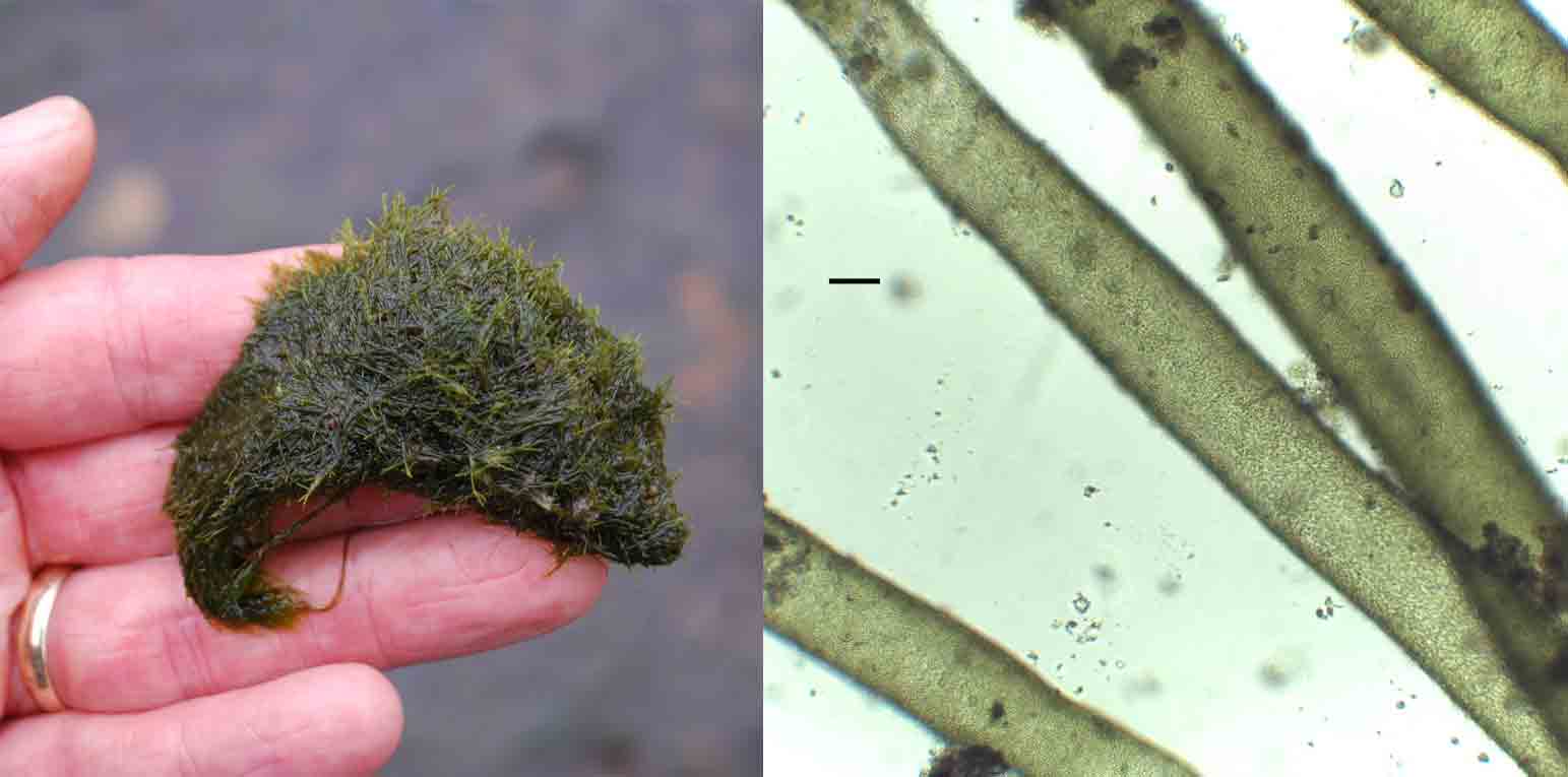
My eye was caught straight away by bright green growths on the shelf on the wall beside the slipway.  Their distinctly felty texture made it easy to recognise the genus as Vaucheria although naming the species is a more difficult job.  We’ve met Vaucheria several times in freshwater (see “the pros and cons of cell walls …”) but it is also commonly found in brackish habitats.  My interest today was not in what species was growing, but in the way that it was helping to build a habitat within which a clump of moss and the shoot of a flowering plant were able to grow.  We saw this with happening with Rhizoclonium riparian during my previous visit (see “If only we looked …”); this time it is an alga from a different phylum, but we are looking at essentially the same phenomenon.

A patch of Vaucheria growing on the wooden training wall beside Deptford Creek, March 2024.  Note the moss (back right) and the shoot that have established themselves in the algal patch.  The patch is about 30 cm across.   The photo at the top of the post shows the training wall beside the slipway at Deptford Creek. 

Underneath this shelf, there was a distinct yellow-brown patch on the wall whose dominant constituent was Hydrosera triquetra.  I commented on its absence in a previous post (see “Deptford’s Heart of Darkness …”) but, in truth, the mudflat is not where I would most expect to find it.  It is a chain-forming diatom that seems to be associated with vertical surfaces and which can tolerate exposure. When individual cells are seen end-on (“valve view”), they are very distinctive, resembling a Star of David, with two overlapping triangles; however, the cells attach at these faces and the characteristic shape is not immediately apparent when seen in fresh material.  You need to focus carefully to understand the third dimension (see “Seeing with my fingers …”).  

Hydrosera triequetra in Deptford Creek, March 2024.  The left-hand image shows a patch growing on a wooden support (the central one visible in the image at the top of the post). The right-hand image shows a magnified view. The scale bar is 20 micrometres (= 1/50th of a millimetre) long.  

Hydrosera triquetra was first observed in the Thames in the early 1970s, and is widely-assumed to have been accidentally introduced, perhaps by a ship discharging ballast water.  Now it is widely-established.  I’m often sceptical of claims about “alien” species  but H. triquetra is sufficiently large and distinctive that it seems unlikely that it would not have been seen by earlier generations of microscopists at least occasionally.  Whether it justifies the term “invasive” is a moot point (see “Pens are too light …”).   That would require some evidence of wholesale changes in both the composition of the microscopic flora, and the way it functioned.  Personally, I love the diversity of modern London and am prepared to embrace a migrant diatom or two without feeling the need to apply pejorative adjectives.  

On the opposite side of the slipway, I noticed a seepage that, again, had a golden-brown hue suggestive of diatoms.  However, I could not see any Hydrosera growing amidst this assemblage, instead, there was a mix of Melosira varians and Biddulphia pulchella, a relative of Hydrosera, which lives in brackish water and forms zigzag colonies.  Motile diatoms such as Nitzschia sigma glided in and around these filaments.  The presence of Melosira varians is interesting.  I am fairly sure, based on examination of living cells, that these filaments are not one of the related species of Melosira that are more tolerant of salinity. That a freshwater species with only mild affinity for salt is thriving here supports some observations of higher plants on the slipway made by Nick and Andy from Creekside Discovery Centre. They noticed that these, too, are mostly not especially salt-tolerant plants, leading them to think that the upper slipway represents a freshwater tidal marsh, rather than a salt marsh, and that it is the result of fresh water from the Ravensbourne being lifted above the denser salt water that pushes into Deptford Creek on each tidal cycle.  A freshwater species tolerant of some salt rubbing along beside a brackish species that can cope with an occasional dose of freshwater seems like a good metaphor for this part of London where people from every part of the world live side-by-side.

Another seepage beside the slipway at Deptford Creek, this time with a mix of Melosira varians (right) and Biddulphia pulchella (left). The scale bar is 20 micrometres (= 1/50th of a millimetre) long.

Some other highlights from this week:

Wrote this whilst listening to:  vintage Jefferson Airplane and, for contrast, Kendrick Lamar’s To Pimp A Butterfly.

Currently reading:  The Secret Diaries of Charles Ignatius Sancho by Patterson Joseph, a novel that is, by coincidence, about 18th century migrants in London.

Cultural highlight:  Lines, a play at the Crucible in Sheffield exploring injustice in Palestine and Uganda.  Rather challenging.

Culinary highlight:  Seven course tasting menu at Prashad, an Indian vegetarian restaurant  in Drighlington, just south of Leeds

When a green alga is not necessarily a Green Alga…

Tribonema_Norfolk_pond_GPhillips

I will end this short series of posts on the organisation of the major groups of algae with a look at the Xanthophyceae, or yellow-green algae.   My old copy of West and Fritsch’s Treatise on British Freshwater Algae from 1927 includes this group of algae with the green algae, although we now know that, apart from a generally green appearance, these two groups of algae have very little in common.  The big differences lie, however, in the types of details that are beyond the purview of the casual natural historian, so you may well find yourself flicking back and forth between “green algae” and “yellow-green algae” as you try to put a name on a specimen.  The definitive test is to add some iodine to your sample, as the Xanthophyceae do not produce starch as a storage product, and so do not produce the characteristic blue-black colour in the cells.  However, iodine is messy stuff and most of us will struggle along without for as long as possible.

The five orders of Xanthophyceae are shown in the table below.   In contrast to the case for most algal groups where molecular studies have led to many revisions of traditional classifications, the Orders of the Xanthophyceae have proved to be quite robust when subjected to this type of scrutiny.   Two of the Orders have siphonous organisation, though the form that this takes is very different in each (see “The pros and cons of cell walls” for more about siphonous lifestyles).  Tribonematales is an Order of filamentous algae that can be difficult to differentiate from filamentous green algae, whilst the Mischococcales are easily confused with small Chlorophyceae.

Xanthophyceae_organisation

The organisation of the Xanthophyceae into five orders.  Organisation follows Algaebase.   The image at the top of this post shows Tribonema smothering the surface of a pond in Norfolk (photo: Geoff Phillips).

That’s one of the mysteries of freshwater algae: to the lay observer, an organism such as Vaucheria looks very similar to Cladophora or another green alga.  Yet they are distant relatives, belonging to different Kingdoms (Chromista and Plantae respectively).  That means that they share the same genetic affinity to one another as they do to us, which is a staggering thought (see “Who do you think you are?”).   What we are seeing is two organisms supremely well adapted to living in similar habitats, which means that natural selection has, gradually, shaped two quite distinct gene pools in quite different ways to arrive at the same end-point.   Just as motor manufacturers have, in the hatchback, found a style of car that is well-adapted to urban living, so the rival algae manufacturing corporations (“Plantae Inc” and “Chromista plc”) have come up with two broadly similar models that are both well-adapted to life in lowland streams.  Just as, in the case of hatchbacks, you can lift up the bonnet and see differences in the engine (petrol, diesel, hybrid, electric) but within the same basic shape, so many of the big differences in algal groups concern their internal machinery not outward appearances.

Vaucheria-frigida_ChrisCarter

Reproductive structures growing from a filament of Vaucheria frigida (photo: Chris Carter)

References

Maistro, S., Broady, P.A., Andreoli, C. & Negrisolo, S. (2009).  Phylogeny and taxonomy of Xanthophyceae (Stramenopiles, Chromalveolata).  Protist 160: 412-426.

Appendix

Links to posts describing representatives of the major groups of Xanthophyceae found in freshwaters.  Only the most recent posts are included, but these should contain links to older posts (you can also use the WordPress search engine to find older posts).

Group Link
Botrydiales Botryidium: The littoral ecology of Lough Down
Mischococcales Watch this space …
Rhizochloridales Watch this space …
Tribonemetales Tribonema: Survival of the fittest (1)
Vaucheriales Vaucheria: When the going gets tough …

Some other highlights from this week:

Wrote this whilst listening to: Two Hands, by Big Thief

Cultural highlights:  Jon Hopkins at the Sage.  What Radio 3’s Ibiza night might sound like.

Currently reading: the last few pages of Bill Bryson’s The Body: A Guide for Occupants (454 pages) prior to starting Hilary Mantel’s The Mirror and The Light (904 pages)

Culinary highlight: fish pie.  Spécialitié de la maison.


The pros and cons of cell walls …

Vaucheria_x100_190630_#1

When I wrote about Vaucheria in a recent post I mentioned that it is “siphonous”, meaning that there are no cross walls dividing the filament into individual cells. Instead, the organism consists of branching tubes containing many separate nuclei and chloroplasts (see “When the going gets tough …”). What I did not do was explain why this might be of benefit to a stream-dwelling organism.

Many scientists over the years have considered the benefits that accrued when simple unicellular organisms banded together to form the first multicellular organisms. They’ve come up with a number of theories, all of which may apply in some cases. However, having accepted that multicellular organisms have a number of advantages over single cells, we have to ask why organisms such as Vaucheria seem to have gone one step further: not only have several cells banded together but they also seem to have lost the cell walls that usually separate the individual units. Most biologists, nurtured in the belief that the cell is the basic unit from which organisms are built, will find Vaucheria’s growth form to be a surprise.

The usefulness of cells, in an evolutionary context, is that this opens the way to specialisation and, over time, to the development of tissues within organisms dedicated to particular tasks. We could think of cells as tiny bags, each containing mixtures of enzymes most relevant to their function. Cells in a leaf, for example, will need to focus on producing enzymes required for photosynthesis whilst those in the trunk will be programmed to produce structural tissues. Cells, in other words, facilitate this division of labour within plants. However, a simple filamentous alga has far fewer needs than a mighty oak tree: the water around them provide both support (so complicated structural tissues are not needed) and a supply of nutrients (removing the need for internal plumbing). If there is less need for specialisation, then there is also less need to invest energy in building cell walls which, despite their advantages, also limit the capacity of cells to share resources. Cells of many algae and all higher plants have channels called “plasmadesmata” which link them together; however, these require energy to function.

If you will excuse a topical, somewhat teleological and rather tortuous metaphor, all plants have the choice of either a “trade deal” (investing energy in plasmadesmata in return for resources from neighbouring cells) or “open borders” (ditching cell walls and sharing resources). For most land plants, the former makes more sense; different lineages of algae, on the other hand, have dabbled in both strategies and Vaucheria represents a successful example of the latter. “Successful”, you sneer, “… it thrives in small clumps in polluted rivers. How is that successful?”. That, if I may say so, is a very terrestrio-centric view of the world: Vaucheria lives in a world where it does not need extensive investment in the tissues that comprise the organisms that are paraded before our eyes on the Living Planet. It has all it needs and neither boasts nor worries about tomorrow.

Vaucheria_x400_190630_#2

Siphon of Vaucheria with a side-branch, collected from the River Browney, Co. Durham and photographed at 400x magnification. Note the absence of cell walls. Scale bar: 20 micrometres (= 1/50th of a millimetre). The photograph at the top of the post shows more siphons, at 100x magnification. Scale bar: 100 micrometres (= 1/10th of a millimetre).

We can find siphonous algae not just in the Xanthophyta (the division to which Vaucheria belongs) but also in some lineages of the green algae, including the important marine genus Caulerpera. Cladophora, no stranger to these posts (see “Summertime blues …”) is siphonocladous, rather than strictly siphonous, meaning that it is divided into large cells, each of which contains many nuclei rather than the single nucleus that is characteristic of most cells throughout the plant and animal kingdoms. Balanced against this, there is a much greater number of multicellular algae that retain cell walls, some of which show considerable differentiation of tissue. Modern land plants arose from this latter largely because of the opportunities this differentiation offered. Being siphonous (or siphonocladous) has proved to be a good strategy under certain circumstances but, in turn, limits the options for the species to extend into new habitats.

In my earlier post I used the metaphor of a “sausage skin” to describe Vaucheria. Most of the interior of the organism is taken up by a vacuole, with the chloroplasts and other cell machinery pressed into a narrow band just inside the cell wall. If you watch closely, you can see the chloroplasts in living Vaucheria moving very slowly – a process called “cytoplasmic streaming”. In very bright light the chloroplasts gather at the sides, so protecting each other from harm (see “Good vibrations under the Suffolk sun …” for another way around this problem). The nuclei and mitochondria (the cell’s “batteries”) can also move around and studies have shown all three move by different mechanisms. Being a siphonous organism offers more prospects for this means of adaptation to local circumstances but, overall, the pros are outweighed by the cons, and there are far more genera of multicellular algae than there of siphonous or siphonocladous algae. Vaucheria and other siphonous algae are clearly very successful in a few habitats but the big picture suggests that being truly multicellular offered organisms far more options in the long term.

References
Coneva, V., & Chitwood, D. H. (2015). Plant architecture without multicellularity: quandaries over patterning and the soma-germline divide in siphonous algae. Frontiers in Plant Science. https://doi.org/10.3389/fpls.2015.00287

Herron, M. D., Borin, J. M., Boswell, J. C., Walker, J., Chen, I.-C. K., Knox, C. A., … Ratcliff, W. C. (2019). De novo origins of multicellularity in response to predation. Scientific Reports. https://doi.org/10.1038/s41598-019-39558-8

Canter-Lund, H. & Lund, J.W.G. (1995). Freshwater Algae: Their Microscopic World Explored. Biopress, Bristol.

Ott, D.W. & Brown, R.M. (1974). Developmental cytology of the genus Vaucheria 1. organisation of the vegetative filament. British Phycological Journal 9: 111 – 126.

Pennisi, E. (2018). The momentous transition to multicellular life may not have been so hard after all. Science, New York. doi:10.1126/science.aau5806

Raven, J.A. (1997). Minireview: multiple origins of plasmadesmata. European Journal of Phycology 32: 95-101.

Vroom, P.S. & C.A. Smith (2001). The challenge of siphonous green algae. American Scientist 89: 525-531.

When the going gets tough …

Castle_Eden_Dene_May19

Two months after the visit I described in the previous post I was back at Castle Eden Dene.    The trees were now in leaf and the floor of the forest was carpeted with wild garlic.   The stream, however, had disappeared below the surface and, once again, I could walk along the channel without getting my feet damp.

Having found a rich crop of diatoms on my last visit when the stream was dry I was intrigued to see what was growing on the stones this time, so I used a toothbrush and some water that I had brought along to scrub a few and collected the dislodged material in my white tray.   I was intrigued to see that the suspension that collected in my tray had a distinct green tinge and, when I got a drop of it under my microscope, found it to be dominated by small green cells.  These were superficially similar to the cells of Desmococcus and Apatococcus that I found on the fence in my garden (see “Little Round Green Things …”) but this is a difficult group with not many clear morphological features with which to distinguish genera so I sent a sample off to Dave John for his opinion.

His view is also that groups such as this are almost impossible to identify unless you grow them in the laboratory or have access to DNA sequencing facilities.   He commented that Desmococcus and Apatococcus both have distinctive 2- or 4-celled packets of cells, which were not common in the Castle Eden Dene sample.  Likely candidates are the generaPleurastrumand Pseudopleurococcus, both of which are subaerial or terrestrial.   Perhaps “Little Round Green Things” is as close as we need to go in this particular instance?

CED_biofilm_May19

A distinctly-green suspension of the biofilm on stones at Castle Eden Dene in May 2019 (left) along with a magnified view showing some of the green cells which dominated the sample (right).  Scale bar: 20 micrometres (1/5thof a millimetre).   

A short distance further on I found some mats of entwined filaments on the tops of stones which also piqued my curiosity.   Under the microscope, and with the addition of a drop of water to rehydrate them, these filaments revealed themselves to belong to Vaucheria (see “Who do you think you are?”).   Technically speaking, Vaucheria is not filamentous but “siphonous”, meaning that there are no cross walls but, instead, the organism consists of branching tubes containing many separate nuclei and chloroplasts.  The cell walls of Vaucheria, however, rupture easily releasing the chloroplasts and giving the appearance of an empty sausage skin. In this case, there are still quite a few chloroplasts but a healthy Vaucheria filament has a uniformly dense green appearance that none of those that I saw in Castle Eden Burn possessed.

There was more than just vegetative filaments of Vaucheria here: scattered amongst them were some larger, spheroid or jar-shaped cells, which are part of Vaucheria’s sexual reproduction apparatus.   I’ve talked before in this blog about how sexual reproduction is relatively rare in the filamentous algae that we find in lakes and streams (see “The perplexing case of the celibate alga …”) and Vaucheria is another case in point.   Put simply, many algae do not bother with sexual reproduction when conditions are favourable and they can grow through simple cell division.   If you subjected a Vaucheria filament to Freudian analysis, it would probably tell you that one outcome of sexual reproduction was a 50% dilution of its unique genotype. So why bother if you don’t have to?  On the other hand, sexual reproduction in these organisms usually results in a zygote with a thick wall that is capable of resisting tough conditions.   The complete absence of water in Castle Eden Burn would be one such circumstance.   To put it another way, when the going gets tough, the algae get frisky.

CED_Vaucheria_mat_May19

Mats of Vaucheria growing on a small boulder in Castle Eden Dene in May 2019.  The picture frame in the left hand image is approximately 30 centimetres. 

CED_Vaucheria_May19

Cell walls of Vaucheria, with a few chloroplasts still present, from Castle Eden Burn, May 2019.  Scale bar: 20 micrometres (= 1/50thof a millimetre). 

However, the structures did not really match any pictures that I could find of oogonia or antheridia in Vaucheria.  I passed my images around some friends, and Gordon Beakes suggested that we might be looking at sporangia of chytrids, a group of fungi that have a string of previous convictions for infecting algae (see “Little bugs have littler bugs upon their backs to bite ‘em ….”).  As I was taking the photographs below, a cloud of tiny spores was released, prompting me to call out “come quickly if you want to see an alga ejaculating” before remembering that we had visitors in the house who might think this a little weird (and not just because I had not yet realised that they were, in fact, fungi).   I even took a video.  I’ll upload it to the Dark Web at some point.  There must be a site for fungal-themed pornography out there, if only I took the time to look…

CED_Vaucheria_oospores

Sporangia of chytrids on Vaucheria filaments from Castle Eden Burn, May 2019.  The one on the right was releasing spores (arrowed) at the time the photograph was taken.   Scale bar: 20 micrometres (= 1/50thof a millimetre).  

Who do you think you are?

As well as the diatom growths, the bed of the River Browney was also covered with skeins of a green-coloured alga which, when I removed it, had a soft, felt-like texture.   This is Vaucheria, a very common constituent of enriched rivers in Britain.   Under the microscope, the filaments can be seen to be long tubes (think of sausage skins) within which there are numerous tiny green chloroplasts.   This is not the first bright green alga that I have written about in this blog but appearances, in the world of algae, can often be deceptive.

Browney_Vaucheria

We often see the evolutionary history of life on earth portrayed as a tree whose branches, representing each of the main groups of organisms, diverge again and again, culminating in “twigs” representing each species.   Following any “twig” back towards the “trunk” links to successively larger groupings of organisms.   So, for example, the species to which we belong, Homo sapiens, has no very close relatives, but is linked at the next level (family) to apes such as the chimpanzee.  Humans and chimps are, in turn, linked to the primates (order) such as gibbons and lemurs, which are part the mammals (class), including elephants and lions, which is part of the chordata (phylum) which links us to fish and reptiles.   Finally, the chordate belong to the Animalia (Kingdom) which links us to flies and slugs, and Animalia is part of the Domain Eukaryophyta, which links us to the rest of life except for bacteria.

We can use this analogy to express the relationship between any two organisms in terms of the number of steps along the tree before we find a common relative.  If we compared Vaucheria to Bulbochaete, which we met on 16 August, another bright green growth from the bed of a river, surprisingly we have to take eight steps (equivalent to comparing humans with plants!).   By contrast, the distance between Bulbochaete and Spirogyra is six steps (humans v fish) and between Vaucheria and Melosira (4 September) is a mere four steps (humans v gibbons).

The message is that the affinities amongst the algae are often not best discerned through seemingly obvious characteristics such as colour and shape but through biochemical composition, similarities in reproduction and the life cycle and in the structure of the DNA.  The other message is that the umbrella term “algae”, usually bit-players in any consideration of life on earth, embraces as much diversity as a typical zoo.   This is too easily forgotten, at least in part because they lack the televisual qualities that lend themselves to wildlife documentaries.   Unfortunately, in the process, we also often lose sight of the importance of algae in ecosystems.

Note

There is no universally agreed system of higher classification (see post of 11 August).  For this exercise I used the tree of life project (www.tolweb.org) as the basis for the classification of animals and Algaebase (www.algaebase.org) for the algae.